File:Philine guineensis (10.1111-zoj.12478) Figure 4.jpg
Jump to navigation
Jump to search
Size of this preview: 424 × 600 pixels. Other resolutions: 170 × 240 pixels | 339 × 480 pixels | 543 × 768 pixels | 724 × 1,024 pixels | 2,128 × 3,010 pixels.
Original file (2,128 × 3,010 pixels, file size: 810 KB, MIME type: image/jpeg)
Captions
Captions
Add a one-line explanation of what this file represents
Summary
[edit]_Figure_4.jpg&action=edit§ion=1)
| Description |
English: Figure 4.
|
| Date | |
| Source | https://dx.doi.org/10.1111%2Fzoj.12478 |
| Author | Malaquias, M.A.E., Ohnheiser, L.T., Oskars, T.R., Willassen, E. 2016. Diversity and systematics of philinid snails (Gastropoda: Cephalaspidea) in West Africa with remarks on the biogeography of the region. Zoological Journal of the Linnean Society |
| Permission (Reusing this file) |
This file is licensed under the Creative Commons Attribution 4.0 International license.
|
File history
Click on a date/time to view the file as it appeared at that time.
| Date/Time | Thumbnail | Dimensions | User | Comment | |
|---|---|---|---|---|---|
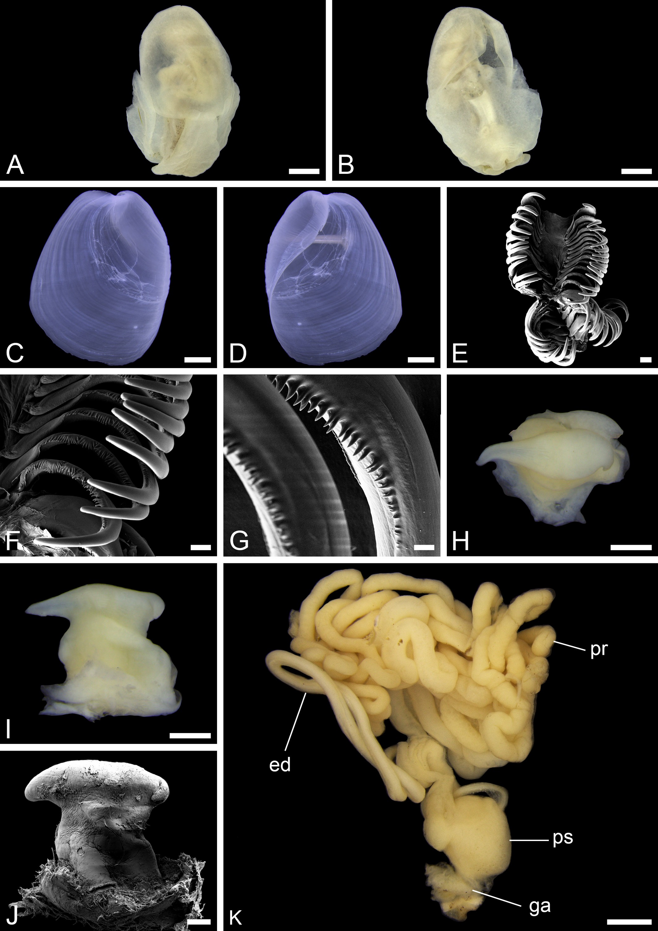
| current | 08:55, 16 July 2021 | | 2,128 × 3,010 (810 KB) | Christian Ferrer (talk | contribs) | {{Information | description = {{en|1=Figure 4. :''Philine guineensis''. A, dorsal view of complete animal. B, ventral view of complete animal. C, dorsal view of shell (automontage image). D, ventral view of shell (automontage image). E, radula (SEM). F, detail of radula (SEM). G, detail of denticulation on inner lateral teeth (SEM). H, top view of penial papilla (automonatge image). I, side view of penial papilla (automontage image). J, side view of penial papilla (SEM). K, male reproductive... |
You cannot overwrite this file.
File usage on Commons
There are no pages that use this file.
_Figure_4.jpg&oldid=702698400)