File:Parasite160090-fig3 Cepedea longa (Opalinidae).png
Jump to navigation
Jump to search

Size of this preview: 610 × 599 pixels. Other resolutions: 244 × 240 pixels | 489 × 480 pixels | 782 × 768 pixels | 1,042 × 1,024 pixels | 1,772 × 1,741 pixels.
Original file (1,772 × 1,741 pixels, file size: 4.44 MB, MIME type: image/png)
Captions
Captions
Add a one-line explanation of what this file represents
Summary
[edit].png&action=edit§ion=1)
| Description |
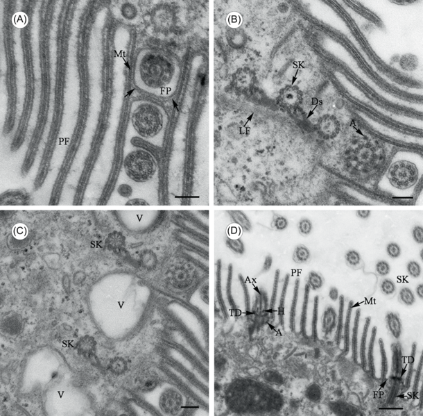
English: Figure 3 of paper Transmission electron microscope images of Cepedea longa, to show fine structures of the somatic flagella.
|
| Date | |
| Source | (2017). "Light and transmission electron microscopy of Cepedea longa (Opalinidae) from Fejervarya limnocharis". Parasite 24: 6. DOI:10.1051/parasite/2017006. ISSN 1776-1042. |
| Author | Can Li, Xiao Jin, Ming Li, Guitang Wang, Hong Zou, Wenxiang Li and Shangong Wu |
Licensing
[edit].png&action=edit§ion=2)
This file is licensed under the Creative Commons Attribution 4.0 International license.
|
 This file was published in the scientific journal Parasite. Their website states that all content of the journal including and after 2013 is published under the Creative Commons Attribution 4.0 license.
|
File history
Click on a date/time to view the file as it appeared at that time.
| Date/Time | Thumbnail | Dimensions | User | Comment | |
|---|---|---|---|---|---|
| current | 10:16, 25 June 2017 | | 1,772 × 1,741 (4.44 MB) | Jeanloujustine (talk | contribs) | User created page with UploadWizard |
You cannot overwrite this file.
File usage on Commons
The following page uses this file:
File usage on other wikis
The following other wikis use this file:
- Usage on en.wikipedia.org
.png&oldid=835646079)