File:Paracentrotus lividus gastrula.jpg
Jump to navigation
Jump to search

Size of this preview: 799 × 599 pixels. Other resolutions: 320 × 240 pixels | 640 × 480 pixels | 1,024 × 768 pixels | 1,245 × 934 pixels.
Original file (1,245 × 934 pixels, file size: 1.08 MB, MIME type: image/jpeg)
Captions
Captions
Add a one-line explanation of what this file represents
Summary
[edit]
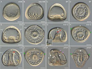
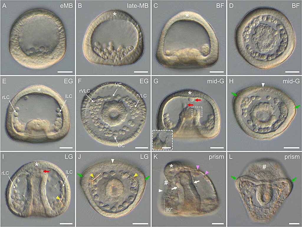
| Description | FIGURE 6. Gastrulation stages of Paracentrotus lividus under light microscopy. Developmental stages are as follows: (A) early mesenchyme blastula stage (eMB); (B) late mesenchyme blastula stage (late-MB); (C,D) blastopore formation stage (BF); (E,F) early gastrula stage (EG); (G,H) mid-gastrula stage (mid-G); (I,J) late gastrula stage (LG); (K,L) prism stage (prism). In (A–C,E,G,I,K), the embryos are in lateral view with the animal pole up, and in (K) the ventral side is left. In (D,F,H,J,L), the embryos are in vegetal view with the ventral side up. In (B,C,E,G,I,K), the asterisk marks the animal (or apical) pole domain. In (G,I), red arrows indicate non-skeletogenic mesoderm cells migrating within the blastocoel. ((G) inset) Close-up of the tip of the archenteron of the same embryo as in (G), but at a different focal plane to illustrate non-skeletogenic mesoderm cell ingression. In (H,J,K), the white arrowhead marks the flattening of the ventral ectoderm. In (H,J,L), green arrows highlight the thickened epithelium at the boundary between the vegetal ventral and the vegetal dorsal ectoderm. In (I,J), yellow arrowheads highlight the presence of skeletal elements. In (K), pink arrowheads indicate red-pigmented cells inserted in the aboral ectoderm, and white arrows mark the constriction of the archenteron segregating the esophagus from the stomach. The white dotted lines with the annotation “90°” further indicate the right angle between the ventral and the vegetal ectoderm. In (K,L), the sign “#” highlights the position of the stomodeum, and thus where the mouth will form. Scale bar: (A–L) 30 μm; ((G) inset) 10 µm. DC: dorsal chain; lLC: left lateral chain; lVLC: left ventrolateral cluster; rLC: right lateral chain; rVLC: right ventrolateral cluster; VC: ventral chain |
| Date | |
| Source |
https://www.frontiersin.org/articles/10.3389/fcell.2022.966408/full Developmental atlas of the indirect-developing sea urchin Paracentrotus lividus: From fertilization to juvenile stages, Front. Cell Dev. Biol., 31 October 2022 Sec. Morphogenesis and Patterning Volume 10 - 2022, https://doi.org/10.3389/fcell.2022.966408 |
| Author | Laurent Formery, Axel Wakefield, Maeva Gesson, Ludovic Toisoul, Guy Lhomond, Laurent Gilletta, Régis Lasbleiz, Michael Schubert, Jenifer C. Croce1 |
Licensing
[edit]
This file is licensed under the Creative Commons Attribution 4.0 International license.
- You are free:
- to share – to copy, distribute and transmit the work
- to remix – to adapt the work
- Under the following conditions:
- attribution – You must give appropriate credit, provide a link to the license, and indicate if changes were made. You may do so in any reasonable manner, but not in any way that suggests the licensor endorses you or your use.

|
This file, which was originally posted to an external website, has not yet been reviewed by an administrator or reviewer to confirm that the above license is valid. See Category:License review needed for further instructions.
|
File history
Click on a date/time to view the file as it appeared at that time.
| Date/Time | Thumbnail | Dimensions | User | Comment | |
|---|---|---|---|---|---|
| current | 01:37, 5 March 2024 | | 1,245 × 934 (1.08 MB) | Rasbak (talk | contribs) | {{Information |description=FIGURE 6. Gastrulation stages of Paracentrotus lividus under light microscopy. Developmental stages are as follows: (A) early mesenchyme blastula stage (eMB); (B) late mesenchyme blastula stage (late-MB); (C,D) blastopore formation stage (BF); (E,F) early gastrula stage (EG); (G,H) mid-gastrula stage (mid-G); (I,J) late gastrula stage (LG); (K,L) prism stage (prism). In (A–C,E,G,I,K), the embryos are in lateral view with the animal pole up, and in (K) the ventral si... |
You cannot overwrite this file.
File usage on Commons
The following 2 pages use this file:
File usage on other wikis
The following other wikis use this file:
- Usage on nl.wikipedia.org