File:PMC4249716 13024 2013 546 Fig7 HTML.png
Jump to navigation
Jump to search
No higher resolution available.
PMC4249716_13024_2013_546_Fig7_HTML.png (512 × 234 pixels, file size: 104 KB, MIME type: image/png)
Captions
Captions
diffuse axonal injury
Summary
[edit]
| Description |
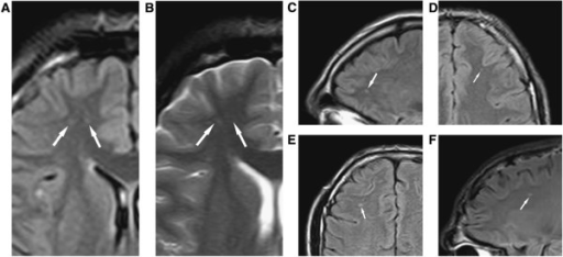
English: Fig7: Diffuse axonal injury on MRI. (Panels A–F) Typical diffuse axonal injury, indicated by arrow. DAI was defined as focal areas of abnormal increased signal intensity on FLAIR and T2-weighted sequences, measuring up to 5 mm in maximum diameter, and located at the gray matter = white matter interface or within or adjacent to the corpus callosum. In the Orrison et al. sample, 29% had DAI. Reproduced Ïrom Orrison et al.[107] with permission. |
| Date | |
| Source | https://openi.nlm.nih.gov/detailedresult?img=PMC4249716_13024_2013_546_Fig7_HTML&query=Diffuse%20axonal%20injury&it=xg&req=4&npos=10 |
| Author | Gandy S, Ikonomovic MD, Mitsis E, Elder G, Ahlers ST, Barth J, Stone JR, DeKosky ST |
Licensing
[edit]
| This file is made available under the Creative Commons CC0 1.0 Universal Public Domain Dedication. | |
| The person who associated a work with this deed has dedicated the work to the public domain by waiving all of their rights to the work worldwide under copyright law, including all related and neighboring rights, to the extent allowed by law. You can copy, modify, distribute and perform the work, even for commercial purposes, all without asking permission.
|

|
This media file is uncategorized.
Please help improve this media file by adding it to one or more categories, so it may be associated with related media files (how?), and so that it can be more easily found.
Please notify the uploader with {{subst:Please link images|File:PMC4249716 13024 2013 546 Fig7 HTML.png}} ~~~~ |
File history
Click on a date/time to view the file as it appeared at that time.
| Date/Time | Thumbnail | Dimensions | User | Comment | |
|---|---|---|---|---|---|
| current | 21:28, 13 August 2023 | | 512 × 234 (104 KB) | Ozzie10aaaa (talk | contribs) | Uploaded a work by Gandy S, Ikonomovic MD, Mitsis E, Elder G, Ahlers ST, Barth J, Stone JR, DeKosky ST from https://openi.nlm.nih.gov/detailedresult?img=PMC4249716_13024_2013_546_Fig7_HTML&query=Diffuse%20axonal%20injury&it=xg&req=4&npos=10 with UploadWizard |
You cannot overwrite this file.
File usage on Commons
There are no pages that use this file.