File:PLoS ONE 0001944 g001 MRS.jpg
Jump to navigation
Jump to search
Size of this preview: 734 × 599 pixels. Other resolutions: 294 × 240 pixels | 588 × 480 pixels | 941 × 768 pixels | 1,254 × 1,024 pixels | 2,048 × 1,672 pixels.
Original file (2,048 × 1,672 pixels, file size: 253 KB, MIME type: image/jpeg)
Captions
Captions
Add a one-line explanation of what this file represents
| Description |
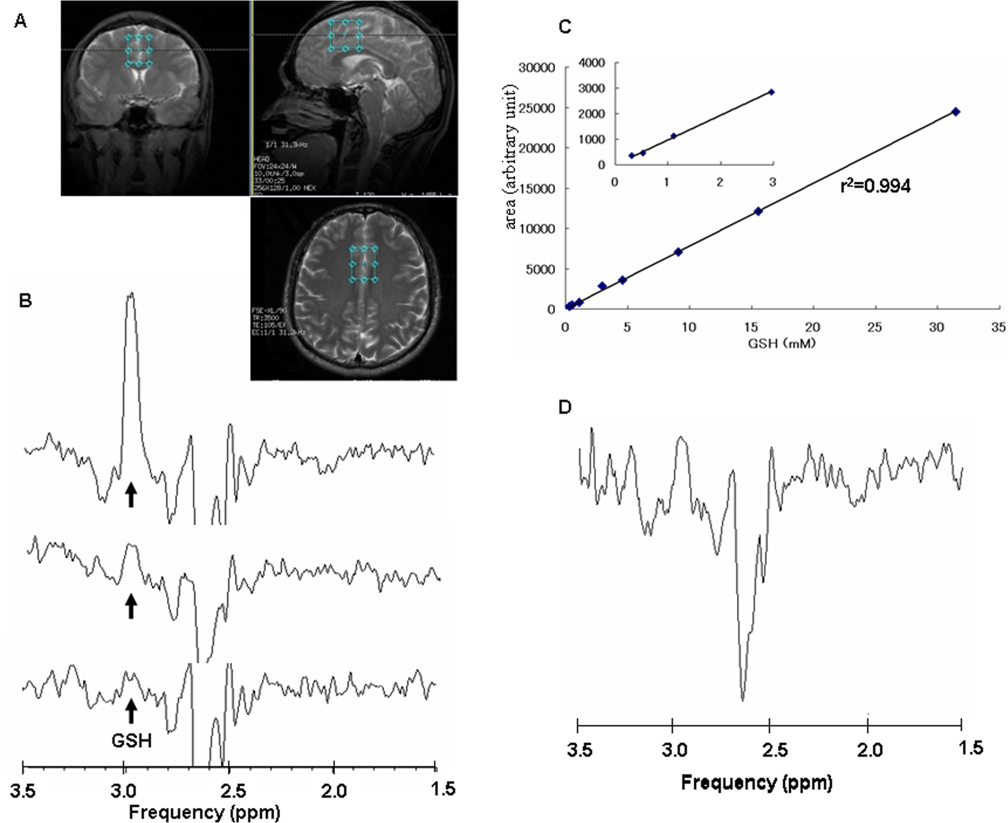
Deutsch: Magnetresonanzspektroskopie eines Abschnittes im Gehirn eines Patienten. Die drei MRT-Aufnahmen zeigen das türkis umrahmte Messgebiet an. Darunter die dazugehörigen NMR-Spektren mit dem Peak von Glutathion (GSH). Rechts oben das daraus abgeleitete Diagramm aus Signal (y-Achse) und Konzentration (x-Achse). |
| Date | |
| Source | Daisuke Matsuzawa, Takayuki Obata, Yukihiko Shirayama, Hiroi Nonaka, Yoko Kanazawa, Eiji Yoshitome, Junichi Takanashi, Tsuyoshi Matsuda, Eiji Shimizu, Hiroo Ikehira, Masaomi Iyo, Kenji Hashimoto: Negative Correlation between Brain Glutathione Level and Negative Symptoms in Schizophrenia: A 3T 1H-MRS Study. In: PLoS ONE 2008 Apr 9;3(4):e1944. doi:10.1371/journal.pone.0001944. PMID 18398470 |
| Author | Daisuke Matsuzawa, Takayuki Obata, Yukihiko Shirayama, Hiroi Nonaka, Yoko Kanazawa, Eiji Yoshitome, Junichi Takanashi, Tsuyoshi Matsuda, Eiji Shimizu, Hiroo Ikehira, Masaomi Iyo, Kenji Hashimoto |
This file is licensed under the Creative Commons Attribution 2.5 Generic license.
- You are free:
- to share – to copy, distribute and transmit the work
- to remix – to adapt the work
- Under the following conditions:
- attribution – You must give appropriate credit, provide a link to the license, and indicate if changes were made. You may do so in any reasonable manner, but not in any way that suggests the licensor endorses you or your use.
File history
Click on a date/time to view the file as it appeared at that time.
| Date/Time | Thumbnail | Dimensions | User | Comment | |
|---|---|---|---|---|---|
| current | 17:07, 19 March 2009 | | 2,048 × 1,672 (253 KB) | Kuebi (talk | contribs) | {{Information |Description= {{de|Magnetresonanzspektroskopie eines Abschnittes im Gehirn eines Patienten. Die drei MRT-Aufnahmen zeigen das türkis umrahmte Messgebiet an. Darunter die dazugehörigen NMR-Spektren mit dem Peaks von Glutathion (GSH). Rechts |
You cannot overwrite this file.
File usage on Commons
The following page uses this file:
File usage on other wikis
The following other wikis use this file:
- Usage on ca.wikipedia.org
- Usage on de.wikipedia.org
- Usage on fr.wikipedia.org