File:Osteochondritis Dissecans lat.-medial-fem.-condyle.jpg
Jump to navigation
Jump to search

Size of this preview: 423 × 599 pixels. Other resolutions: 169 × 240 pixels | 497 × 704 pixels.
Original file (497 × 704 pixels, file size: 138 KB, MIME type: image/jpeg)
Captions
Captions
Add a one-line explanation of what this file represents
Summary
[edit]
| Description |
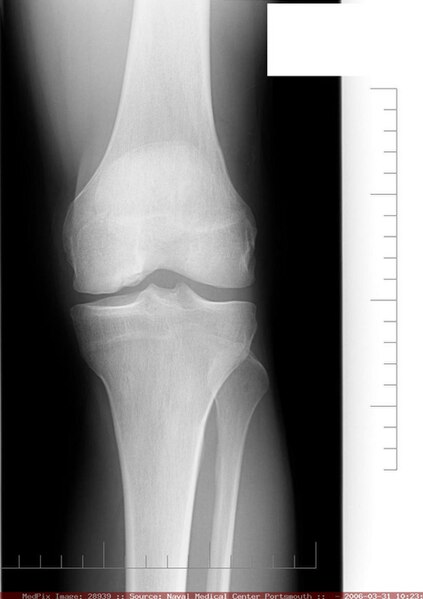
English: "There is fragmentation of the lateral aspect of the medial femoral condyle consistent with osteochondritis dissecans. There are two ossific fragments present. The largest measuring 0.9 cm, the smaller measuring 0.4 cm. No evidence of intra-articular body. No evidence of effusion." |
| Date | |
| Source |
Naval Medical Center Portsmouth Obtained from MedPix Database: http://rad.usuhs.edu/medpix/medpix_image.html?imageid=28939 |
| Author |
Author: Kenneth T Blackner, Affiliation: Naval Medical Center Portsmouth Editor: Stephanie A Bernard, Affiliation: Naval Medical Center Portsmouth |
History of left knee pain prior to plain film imaging.
Licensing
[edit]
This file is a work of a sailor or employee of the U.S. Navy, taken or made as part of that person's official duties. As a work of the U.S. federal government, it is in the public domain in the United States.
|
 | |
| This file has been identified as being free of known restrictions under copyright law, including all related and neighboring rights. | ||
File history
Click on a date/time to view the file as it appeared at that time.
| Date/Time | Thumbnail | Dimensions | User | Comment | |
|---|---|---|---|---|---|
| current | 23:30, 10 October 2008 | | 497 × 704 (138 KB) | FoodPuma (talk | contribs) | {{Information |Description={{en|1="There is fragmentation of the lateral aspect of the medial femoral condyle consistent with osteochondritis dissecans. There are two ossific fragments present. The largest measuring 0.9 cm, the smaller measuring 0.4 cm. N |
You cannot overwrite this file.
File usage on Commons
There are no pages that use this file.
File usage on other wikis
The following other wikis use this file:
- Usage on en.wikipedia.org