File:Origin of Vertebrates Fig 058.png
Jump to navigation
Jump to search

Size of this preview: 546 × 599 pixels. Other resolutions: 219 × 240 pixels | 437 × 480 pixels | 700 × 768 pixels | 933 × 1,024 pixels | 1,668 × 1,831 pixels.
Original file (1,668 × 1,831 pixels, file size: 395 KB, MIME type: image/png)
Captions
Captions
Add a one-line explanation of what this file represents
Summary
[edit]
| Description |
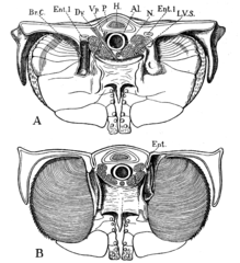
English: Fig. 58.—Transverse Section through the Mesosoma of Limulus, to show the Anterior (A) and the Posterior (B) Surfaces of a Mesosomatic or Branchial Appendage.
|
| Date | |
| Source | The Origin of Vertebrates. https://archive.org/details/originofvertebra1908gask |
| Author | Walter Holbrook Gaskell. |
Licensing
[edit]
|
The author died in 1914, so this work is in the public domain in its country of origin and other countries and areas where the copyright term is the author's life plus 100 years or fewer. This work is in the public domain in the United States because it was published (or registered with the U.S. Copyright Office) before January 1, 1929. | |
| This file has been identified as being free of known restrictions under copyright law, including all related and neighboring rights. | |
File history
Click on a date/time to view the file as it appeared at that time.
| Date/Time | Thumbnail | Dimensions | User | Comment | |
|---|---|---|---|---|---|
| current | 14:08, 10 November 2013 | | 1,668 × 1,831 (395 KB) | Keith Edkins (talk | contribs) | == {{int:filedesc}} == {{Information |Description={{En|Fig. 58.—Transverse Section through the Mesosoma of Limulus, to show the Anterior (A) and the Posterior (B) Surfaces of a Mesosomatic or Branchial Appendage. In each figure the branchial cartil... |
You cannot overwrite this file.
File usage on Commons
There are no pages that use this file.
File usage on other wikis
The following other wikis use this file: