File:Ophiuroglypha fendouzhe (10.5852-ejt.2023.891.2281) Figure 4.png
Jump to navigation
Jump to search

Size of this preview: 485 × 599 pixels. Other resolutions: 194 × 240 pixels | 389 × 480 pixels | 622 × 768 pixels | 1,264 × 1,561 pixels.
Original file (1,264 × 1,561 pixels, file size: 793 KB, MIME type: image/png)
Captions
Captions
Add a one-line explanation of what this file represents
Summary
[edit]_Figure_4.png&action=edit§ion=1)
| Description |
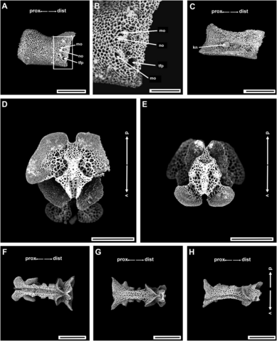
English: Fig. 4. Ophiuroglypha fendouzhe sp. nov., paratype (IDSSE-EEB-SW0251). A–C. Lateral arm plate. D–H. Vertebrae. D. Distal view (right side of the structure partially broken). E. Proximal view. F. Ventral view. G. Dorsal view. H. Dorsolateral view. Abbreviations: asa = arm spine articulation; d = dorsal; dist = distal; kn = knob; mo = muscle opening; no = nerve opening; pb = podial basin; prox = proximal; tfp = tube foot pore; v = ventral. Scale bars: A, C, F–H = 500 μm; B = 100 μm; D–E = 300 μm. |
| Date | |
| Source | Nethupul, H., Stöhr, S., & Zhang, H. (2023). Deepest known novel species of the genus Ophiuroglypha Hertz, 1927 (Echinodermata: Ophiuroidea) from the central rift zone, Philippine Sea. European Journal of Taxonomy, 891(1), 167-185. https://doi.org/10.5852/ejt.2023.891.2281 |
| Author | Nethupul, H., Stöhr, S., & Zhang, H. (2023) |
| Permission (Reusing this file) |
This file is licensed under the Creative Commons Attribution 4.0 International license.
|
File history
Click on a date/time to view the file as it appeared at that time.
| Date/Time | Thumbnail | Dimensions | User | Comment | |
|---|---|---|---|---|---|
| current | 11:28, 5 October 2023 | | 1,264 × 1,561 (793 KB) | Christian Ferrer (talk | contribs) | {{Information | description = {{en|1=Fig. 4. ''Ophiuroglypha fendouzhe'' sp. nov., paratype (IDSSE-EEB-SW0251). A–C. Lateral arm plate. D–H. Vertebrae. D. Distal view (right side of the structure partially broken). E. Proximal view. F. Ventral view. G. Dorsal view. H. Dorsolateral view. Abbreviations: asa = arm spine articulation; d = dorsal; dist = distal; kn = knob; mo = muscle opening; no = nerve opening; pb = podial basin; prox = proximal; tfp = tube foot pore; v = ventral. Scale bars: A... |
You cannot overwrite this file.
File usage on Commons
The following page uses this file:
_Figure_4.png&oldid=903973309)