File:Oby007f8b.png
Jump to navigation
Jump to search

Size of this preview: 431 × 599 pixels. Other resolutions: 172 × 240 pixels | 345 × 480 pixels | 552 × 768 pixels | 736 × 1,024 pixels | 1,700 × 2,364 pixels.
Original file (1,700 × 2,364 pixels, file size: 7.86 MB, MIME type: image/png)
Captions
Captions
Add a one-line explanation of what this file represents
Summary
[edit]
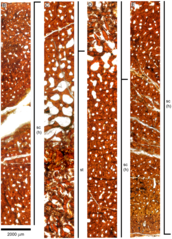
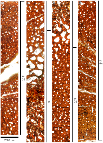
| Description | Overview (A) and microanatomy (B–E) of the adult D. bonneri humerus, UNSM 50133. Black boxes indicate magnified regions in this figure and Fig. 11. The secondary trabeculae in the center of the element have collapsed due to brittle deformation of the humerus after burial. Note lack of EFS, unlike the Polycotylus mother. a, anterior; d, dorsal; p, posterior; sc(h), secondary cortex, Haversian; st, secondary trabeculae; v, ventral. |
| Date | |
| Source | F R O’Keefe, P M Sander, T Wintrich, S Werning; Ontogeny of Polycotylid Long Bone Microanatomy and Histology, Integrative Organismal Biology, Volume 1, Issue 1, 1 January 2019, oby007, https://doi.org/10.1093/iob/oby007 |
| Author | F R O’Keefe P M Sander T Wintrich S Werning |
Licensing
[edit]
This file is licensed under the Creative Commons Attribution 4.0 International license.
- You are free:
- to share – to copy, distribute and transmit the work
- to remix – to adapt the work
- Under the following conditions:
- attribution – You must give appropriate credit, provide a link to the license, and indicate if changes were made. You may do so in any reasonable manner, but not in any way that suggests the licensor endorses you or your use.
File history
Click on a date/time to view the file as it appeared at that time.
| Date/Time | Thumbnail | Dimensions | User | Comment | |
|---|---|---|---|---|---|
| current | 18:09, 29 January 2019 | | 1,700 × 2,364 (7.86 MB) | Abyssal (talk | contribs) | User created page with UploadWizard |
You cannot overwrite this file.
File usage on Commons
There are no pages that use this file.