File:Morbus Fabry Skin 01.jpg
From Wikimedia Commons, the free media repository
Jump to navigation
Jump to search

Size of this preview: 800 × 575 pixels. Other resolutions: 320 × 230 pixels | 640 × 460 pixels | 1,024 × 736 pixels | 1,200 × 862 pixels.
Original file (1,200 × 862 pixels, file size: 128 KB, MIME type: image/jpeg)
File information
Structured data
Captions
Captions
Add a one-line explanation of what this file represents
Morbus Fabry Skin 01.jpg
| DescriptionMorbus Fabry Skin 01.jpg |
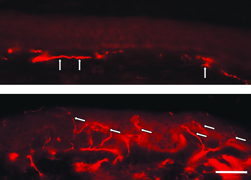
English: Photomicrographs of frozen skin sections (50 µm) from a Fabry patient. Samples immunoreacted with PGP 9.5 and were processed for fluorescence microscopy with Cy3 labelled secondary antibodies. Note the lack of intraepidermal nerve fibers and persistence of fibers pertaining to the subepidermal nerve plexus (arrows) in the sample from the lower leg skin of a Fabry patient (upper figure). Note the dense innervation of the epidermis (arrows) in the sample from the back of the Fabry patient, taken at the dermatome Th 12 (lower figure). Bar eq. 50 µm.
Deutsch: Fluoreszenzmikroskopaufnahmen eines Gefrierschnittes des Haut eines Morbus-Fabry-Patienten. Die Hautprobe des Unterschenkels (obere Aufnahme) wurde mit einem PGP-9.5-Antikörper versetzt und mit einem zweiten, mit dem Fluoreszenzfarbstoff Cy3 markierten, Antikörper versetzt. Auffällig ist der Mangel an intraepidermalen Nervenfasern und das Vorhandensein von Fasern, die zum subepidermalen Nervenplexus gehören (Pfeile). Die untere Hautprobe stammt dagegen vom Rücken des Patienten. Hier ist die dichte Innervation der Epidermis (Pfeile) auffällig. Die Länge des weißen Balkens in der Aufnahme betägt 50 µm. Die Proben wurden mit einem Dermatom entnommen. |
| Date | article published: 27 May 2011 [1] |
| Source | Alessandro P Burlina, Katherine B Sims, Juan M Politei, Gary J Bennett, Ralf Baron, Claudia Sommer, Anette Torvin Møller and Max J Hilz: Early diagnosis of peripheral nervous system involvement in Fabry disease and treatment of neuropathic pain: the report of an expert panel. In: BMC Neurology 2011, 11:61 doi:10.1186/1471-2377-11-61 |
| Author | Alessandro P Burlina, Katherine B Sims, Juan M Politei, Gary J Bennett, Ralf Baron, Claudia Sommer, Anette Torvin Møller and Max J Hilz |
This file is licensed under the Creative Commons Attribution 2.0 Generic license.
- You are free:
- to share – to copy, distribute and transmit the work
- to remix – to adapt the work
- Under the following conditions:
- attribution – You must give appropriate credit, provide a link to the license, and indicate if changes were made. You may do so in any reasonable manner, but not in any way that suggests the licensor endorses you or your use.
File history
Click on a date/time to view the file as it appeared at that time.
| Date/Time | Thumbnail | Dimensions | User | Comment | |
|---|---|---|---|---|---|
| current | 19:33, 31 August 2011 |  | 1,200 × 862 (128 KB) | Kuebi (talk | contribs) | Morbus Fabry Skin 01.jpg {{Information |Description={{en|Photomicrographs of frozen skin sections (50 µm) from a Fabry patient. Samples immunoreacted with PGP 9.5 and were processed for fluorescence microscopy with Cy3 labelled secondary antibodies. Note |
You cannot overwrite this file.
File usage on Commons
The following 2 pages use this file:
File usage on other wikis
The following other wikis use this file:
- Usage on de.wikipedia.org
- Usage on or.wikipedia.org
- Usage on outreach.wikimedia.org
- Usage on sr.wikipedia.org
Metadata
This file contains additional information such as Exif metadata which may have been added by the digital camera, scanner, or software program used to create or digitize it. If the file has been modified from its original state, some details such as the timestamp may not fully reflect those of the original file. The timestamp is only as accurate as the clock in the camera, and it may be completely wrong.
| _error | 0 |
|---|