File:Models of SMC and cohesin structure.svg
Jump to navigation
Jump to search

Size of this PNG preview of this SVG file: 234 × 598 pixels. Other resolutions: 94 × 240 pixels | 188 × 480 pixels | 300 × 768 pixels | 401 × 1,024 pixels | 801 × 2,048 pixels | 512 × 1,308 pixels.
Original file (SVG file, nominally 512 × 1,308 pixels, file size: 571 KB)
Captions
Captions
Models of SMC and cohesin structure
Summary
[edit]
| Description |
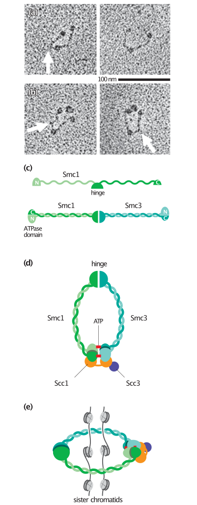
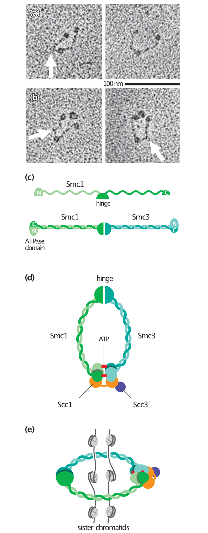
English: a) Electron microscopy of Xenopus Smc1–Smc3 dimers illustrates the V-shaped structure commonly seen with SMC proteins. The flexible ‘hinge’ region is at the bottom of the V and the two globular ATPase domains are at the top. The arrow indicates a kink that is sometimes seen in one arm. (b) Addition of the two non-SMC subunits (Scc1 and Scc3) of the cohesin complex results in the appearance of a globular structure next to the two heads of the Smc1–Smc3 dimer. (c) The linear structure of an SMC protein includes two globular domains at each terminus, linked by a long repetitive sequence and a central dimerization or hinge domain. When the SMC protein is folded, the two domains at the termini join to form a complete ATPase domain, while the arm regions form a helical coiled-coil. The hinge domain that forms at the other end of the arm interacts with the hinge domain of another SMC protein. In cohesin, this results in the formation of a Smc1–Smc3 heterodimer. (d) Binding of ATP (red) promotes binding of the two ATPase domains, resulting in closure of the SMC ring. The non-SMC protein Scc1 interacts with both ATPase domains and holds them together. Cleavage of Scc1 in anaphase therefore opens the ring. (e) The cohesin complex may form a 50-nm ring around two sister chromatids. Because of its small size, however, this ring could only link nucleosomal DNA and not more complex chromatin structures. Panels (a) and (b) from Anderson, D.E. et al.: J. Cell Biol. 2002, 156:419–424.[1] |
| Date | |
| Source | The Cell Cycle. Principles of Control. |
| Author | David O Morgan |
Licensing
[edit]

|
The copyright holder of this file allows anyone to use it for any purpose, provided that the copyright holder is properly attributed. Redistribution, derivative work, commercial use, and all other use is permitted. |
|
|
File history
Click on a date/time to view the file as it appeared at that time.
| Date/Time | Thumbnail | Dimensions | User | Comment | |
|---|---|---|---|---|---|
| current | 22:18, 6 May 2020 | 512 × 1,308 (571 KB) | Rob Hurt (talk | contribs) | Uploaded a work by David O Morgan from The Cell Cycle. Principles of Control. with UploadWizard |
You cannot overwrite this file.
File usage on Commons
There are no pages that use this file.
File usage on other wikis
The following other wikis use this file:
- Usage on en.wikipedia.org
- Usage on es.wikipedia.org
- Usage on gl.wikipedia.org
- Usage on ru.wikipedia.org