File:Micrograph of colorectal choriocarcinoma, rhabdoid colorectal carcinoma, carcinoma with osseous metaplasia and undifferentiated carcinoma.jpg
Jump to navigation
Jump to search

Size of this preview: 794 × 600 pixels. Other resolutions: 318 × 240 pixels | 636 × 480 pixels | 1,017 × 768 pixels | 1,280 × 967 pixels | 1,866 × 1,409 pixels.
Original file (1,866 × 1,409 pixels, file size: 1.11 MB, MIME type: image/jpeg)
Captions
Captions
Micrograph of colorectal choriocarcinoma, rhabdoid colorectal carcinoma, carcinoma with osseous metaplasia and undifferentiated carcinoma
Summary
[edit]| Description |
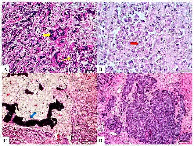
English: Original caption: "Haematoxylin and Eosin stained sections of rare type colorectal carcinomas. (A) Colorectal Choriocarcinoma: biphasic solid nests and trabeculae of mononucleated cells with clear cytoplasm (thin yellow arrow) and pleomorphic cells with abundant vacuolated or eosinophilic cytoplasm and single or multiple vescicular nuclei with conspicuous nucleoli (thick yellow arrow). Scale bar 200 micron. (B) Rhabdoid Colorectal Carcinoma: rhabdoid cells characterized by a large, eccentrically located nuclei, prominent nucleoli (red arrow) and abundant eosinophilic cytoplasm. Scale bar 100 micron. (C) Carcinoma with osseous metaplasia: osseous metaplasia (blue arrow) is recognized in conventional CRC as foci of bone formation in the stroma, with calcification, osteoid matrix, osteoclasts and osteoblasts. Scale bar 400 micron. (D) Undifferentiated carcinoma: sheets of undifferentiated cells showing a variable grade of pleomorphism with no gland formation, mucin production or other line of differentiation. Scale bar 400 micron." |
| Date | |
| Source |
(2019). "Morphology and Molecular Features of Rare Colorectal Carcinoma Histotypes". Cancers 11 (7): 1036. DOI:10.3390/cancers11071036. ISSN 2072-6694. |
| Author | Andrea Remo, Matteo Fassan, Alessandro Vanoli, Luca Reggiani Bonetti, Valeria Barresi, Fabiana Tatangelo, Roberta Gafà, Guido Giordano, Massimo Pancione, Federica Grillo and Luca Mastracci |
| Other versions |
|


Licensing
[edit]
This file is licensed under the Creative Commons Attribution 4.0 International license.
- You are free:
- to share – to copy, distribute and transmit the work
- to remix – to adapt the work
- Under the following conditions:
- attribution – You must give appropriate credit, provide a link to the license, and indicate if changes were made. You may do so in any reasonable manner, but not in any way that suggests the licensor endorses you or your use.
File history
Click on a date/time to view the file as it appeared at that time.
| Date/Time | Thumbnail | Dimensions | User | Comment | |
|---|---|---|---|---|---|
| current | 12:48, 30 September 2019 | | 1,866 × 1,409 (1.11 MB) | Mikael Häggström (talk | contribs) | User created page with UploadWizard |
You cannot overwrite this file.
File usage on Commons
The following 4 pages use this file:
- File:Micrograph of colorectal choriocarcinoma, rhabdoid colorectal carcinoma, carcinoma with osseous metaplasia and undifferentiated carcinoma.jpg
- File:Micrograph of lymphoepitelioma-like carcinoma, cribiform comedo-type carcinoma, micropapillary carcinoma and low grade tubulo-glandular carcinoma.jpg
- File:Micrograph of serrated adenocarcinoma, mucinous carcinoma, signet ring carcinoma and medullary carcinoma.jpg
- Template:Colorectal cancer histopathology types
File usage on other wikis
The following other wikis use this file:
- Usage on en.wikipedia.org