File:Mesenchyme (01).jpg
Jump to navigation
Jump to search

Size of this preview: 800 × 573 pixels. Other resolutions: 320 × 229 pixels | 640 × 458 pixels | 856 × 613 pixels.
Original file (856 × 613 pixels, file size: 271 KB, MIME type: image/jpeg)
Captions
Captions
Add a one-line explanation of what this file represents
Summary
[edit].jpg&action=edit§ion=1)
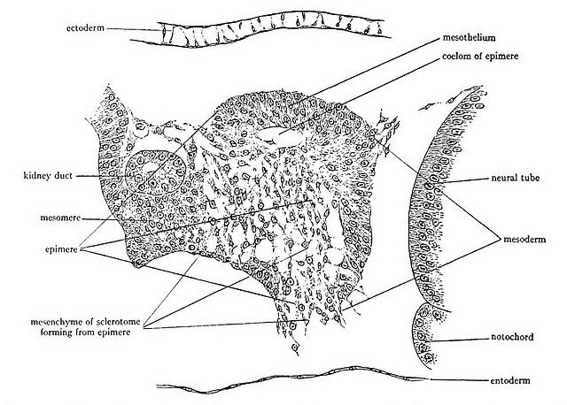
| Description | Fig. 9.—Enlarged view of the epimere (the dorsal part of a mesodermal segment of a chordate embryo) of a chick embryo of two days’ incubation to show the transformation of a portion of the epimere into mesenchyme. These mesenchyme cells constitute the sclerotome from which the vertebral column arises. |
| Date | |
| Source | https://archive.org/details/cu31924021952902/page/42/mode/1up?view=theater A laboratory manual for comparative vertebrate anatomy |
| Author | Hyman, Libbie Henrietta, 1888-1969 |
Licensing
[edit].jpg&action=edit§ion=2)
|
This work is in the public domain in its country of origin and other countries and areas where the copyright term is the author's life plus 70 years or fewer. | |
| This file has been identified as being free of known restrictions under copyright law, including all related and neighboring rights. | |
File history
Click on a date/time to view the file as it appeared at that time.
| Date/Time | Thumbnail | Dimensions | User | Comment | |
|---|---|---|---|---|---|
| current | 01:17, 30 January 2024 | | 856 × 613 (271 KB) | Rasbak (talk | contribs) | {{Information |description=Fig. 9.—Enlarged view of the epimere (the dorsal part of a mesodermal segment of a chordate embryo) of a chick embryo of two days’ incubation to show the transformation of a portion of the epimere into mesenchyme. These mesenchyme cells constitute the sclerotome from which the vertebral column arises. |date=1922 |source=https://archive.org/details/cu31924021952902/page/42/mode/1up?view=theater A laboratory manual for comparative vertebrate anatomy |author=Hyman, Lib... |
You cannot overwrite this file.
File usage on Commons
There are no pages that use this file.
File usage on other wikis
The following other wikis use this file:
.jpg&oldid=846975639)