File:Marburg virus.jpg
Jump to navigation
Jump to search
No higher resolution available.
Marburg_virus.jpg (700 × 474 pixels, file size: 34 KB, MIME type: image/jpeg)
Captions
Captions
Add a one-line explanation of what this file represents
Summary
[edit]| Description |
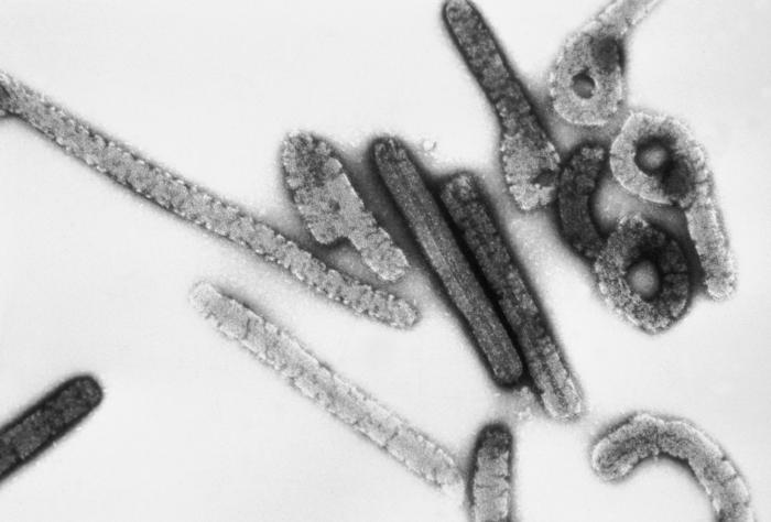
English: This negative stained transmission electron micrograph (TEM) depicts a number of filamentous Marburg virions, which had been cultured on Vero cell cultures, and purified on sucrose, rate-zonal gradients. Note the virus’s morphologic appearance with its characteristic “Shepherd’s Crook” shape; Magnified approximately 100,000x.
Marburg hemorrhagic fever is a rare, severe type of hemorrhagic fever which affects both humans and non-human primates. Caused by a genetically unique zoonotic (that is, animal-borne) RNA virus of the filovirus family, its recognition led to the creation of this virus family. The four species of Ebola virus are the only other known members of the filovirus family. Marburg virus was first recognized in 1967, when outbreaks of hemorrhagic fever occurred simultaneously in laboratories in Marburg and Frankfurt, Germany and in Belgrade, Yugoslavia (now Serbia).
Deutsch: Marburg-Virus.
Français : Vue de particules virales de Marburg au microscope électronique ; on voit la structure typique des filovirus, ainsi que les filaments caractéristiques en forme de crochets. Agrandissement 100 000x.
Polski: Fotografia z elektronowego mikroskopu transmisyjnego ukazująca wiriony wirusa Marburg. Wirus Marburg.
Svenska: Marburgvirus |
||
| Date | |||
| Source |
|
||
| Author |
|
||
| Permission (Reusing this file) |
PD-USGov-HHS-CDC English: None - This image is in the public domain and thus free of any copyright restrictions. As a matter of courtesy we request that the content provider be credited and notified in any public or private usage of this image. |
||
| Other versions |
|

Licensing
[edit]
This image is a work of the Centers for Disease Control and Prevention, part of the United States Department of Health and Human Services, taken or made as part of an employee's official duties. As a work of the U.S. federal government, the image is in the public domain.
|
File history
Click on a date/time to view the file as it appeared at that time.
| Date/Time | Thumbnail | Dimensions | User | Comment | |
|---|---|---|---|---|---|
| current | 23:29, 30 November 2007 | | 700 × 474 (34 KB) | Tijuana Brass~commonswiki (talk | contribs) | Transmission electron micrograph of the Marburg virus. Dr. Erskine Palmer, Russell Regnery, Ph.D., 1981. Public domain. Available online in higher resolution at [http://phil.cdc.gov/phil/home.asp Public Health Image Library], ID# 275. {{Template:PD-USGov |
| 22:37, 20 May 2005 |  | 500 × 397 (18 KB) | NGerda~commonswiki (talk | contribs) | From the CDC. {{PD-USGov}} |
You cannot overwrite this file.
File usage on Commons
The following 6 pages use this file:
File usage on other wikis
The following other wikis use this file:
- Usage on ar.wikipedia.org
- Usage on arz.wikipedia.org
- Usage on azb.wikipedia.org
- Usage on bg.wikipedia.org
- Usage on ca.wikipedia.org
- Usage on cs.wikipedia.org
- Usage on da.wikipedia.org
- Usage on de.wikipedia.org
- Usage on de.wikibooks.org
- Usage on en.wikipedia.org
- Usage on en.wikinews.org
- Usage on en.wikiversity.org
- Usage on en.wiktionary.org
- Usage on es.wikipedia.org
- Usage on et.wikipedia.org
- Usage on eu.wikipedia.org
- Usage on fa.wikipedia.org
View more global usage of this file.