File:Mammals Embryo Origin of the germ layers of mammals as seen in transverse section.jpg
Original file (1,750 × 742 pixels, file size: 422 KB, MIME type: image/jpeg)
Captions
Captions
Summary
[edit]
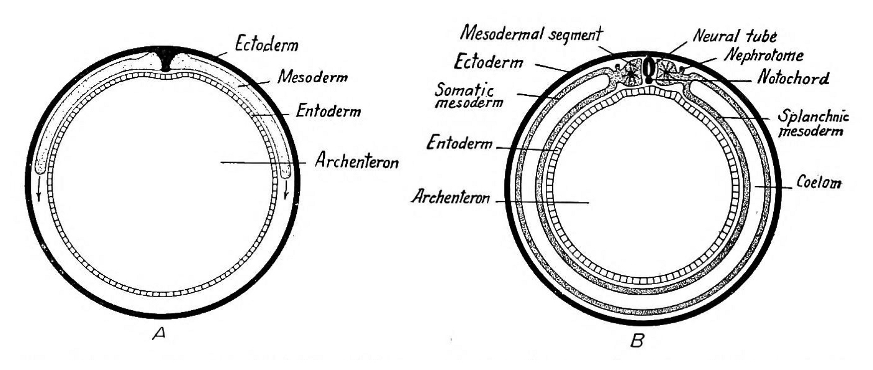
| Description | Fig. 30.—Diagrams showing the origin of the germ layers of mammals as seen in transverse section (modified from Bryce). The mesoderm rapidly grows round the wall of the blastodermic vesicle (A) until it finally surrounds it and the two wings fuse ventrally (B). |
| Date | |
| Source | https://archive.org/details/cu31924001033459/page/46/mode/1up?view=theater A laboratory manual and text-book of embryology. Philadelphia, London, W. B. Saunders. |
| Author | Prentiss, Charles William |
Licensing
[edit]
|
This work is in the public domain in its country of origin and other countries and areas where the copyright term is the author's life plus 70 years or fewer.
| |
| This file has been identified as being free of known restrictions under copyright law, including all related and neighboring rights. | |

|
This file, which was originally posted to an external website, has not yet been reviewed by an administrator or reviewer to confirm that the above license is valid. See Category:License review needed for further instructions.
|
File history
Click on a date/time to view the file as it appeared at that time.
| Date/Time | Thumbnail | Dimensions | User | Comment | |
|---|---|---|---|---|---|
| current | 11:27, 16 March 2024 | | 1,750 × 742 (422 KB) | Rasbak (talk | contribs) | {{Information |description=Fig. 31.—Dorsal view of a twenty-five-hour chick embryo with seven primitive segments. X 20. |date=1915 |source=https://archive.org/details/cu31924001033459/page/49/mode/1up?view=theater A laboratory manual and text-book of embryology. Philadelphia, London, W. B. Saunders. |author= Prentiss, Charles William |other versions= }} =={{int:license-header}}== {{PD-Old}} {{Licensereview}} Category:Endoderm Category:Ectoderm Category:Mesooderm [[Category:Arche... |
You cannot overwrite this file.
File usage on Commons
The following page uses this file: