File:Macular corneal dystrophy hale colloidal iron stain.JPEG
Jump to navigation
Jump to search
No higher resolution available.
Macular_corneal_dystrophy_hale_colloidal_iron_stain.JPEG (500 × 500 pixels, file size: 152 KB, MIME type: image/jpeg)
Captions
Captions
Add a one-line explanation of what this file represents
| Description |
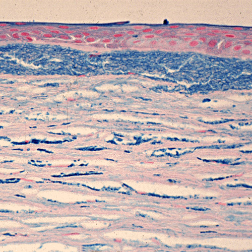
English: Macular corneal dystrophy. The abnormalities within the cornea are easily seen within the keratocytes and in a subepithelial extracellular location because they stain prominently with methods that demonstrate glycosaminoglycans. Hale colloidal iron stain. Klintworth Orphanet Journal of Rare Diseases 2009 4:7 doi:10.1186/1750-1172-4-7 Русский: Пятнистая дистрофия роговицы. Окрашивание гликозаминогликанов выявляет нарушения в кератоцитах стромы и во внеклеточном пространстве под эпителием роговицы. |
| Date | |
| Source | Corneal dystrophies |
| Author | Klintworth GK. |
| Permission (Reusing this file) |
© 2009 Klintworth; licensee BioMed Central Ltd. This is an Open Access article distributed under the terms of the Creative Commons Attribution License (https://creativecommons.org/licenses/by/2.0), which permits unrestricted use, distribution, and reproduction in any medium, provided the original work is properly cited. |
This file is licensed under the Creative Commons Attribution 2.0 Generic license.
- You are free:
- to share – to copy, distribute and transmit the work
- to remix – to adapt the work
- Under the following conditions:
- attribution – You must give appropriate credit, provide a link to the license, and indicate if changes were made. You may do so in any reasonable manner, but not in any way that suggests the licensor endorses you or your use.
File history
Click on a date/time to view the file as it appeared at that time.
| Date/Time | Thumbnail | Dimensions | User | Comment | |
|---|---|---|---|---|---|
| current | 06:42, 5 July 2009 | | 500 × 500 (152 KB) | CopperKettle (talk | contribs) | {{Information |Description={{en|1=Macular corneal dystrophy. The abnormalities within the cornea are easily seen within the keratocytes and in a subepithelial extracellular location because they stain prominently with methods that demonstrate glycosaminog |
You cannot overwrite this file.
File usage on Commons
The following page uses this file:
File usage on other wikis
The following other wikis use this file:
- Usage on de.wikipedia.org
- Usage on en.wikipedia.org
- Usage on es.wikipedia.org
- Usage on it.wikipedia.org
- Usage on ru.wikipedia.org
- Usage on tt.wikipedia.org
- Usage on www.wikidata.org