File:Lophosteus superbus odontodes.jpg
Jump to navigation
Jump to search

Size of this preview: 440 × 600 pixels. Other resolutions: 176 × 240 pixels | 352 × 480 pixels | 563 × 768 pixels | 751 × 1,024 pixels | 1,654 × 2,254 pixels.
Original file (1,654 × 2,254 pixels, file size: 730 KB, MIME type: image/jpeg)
Captions
Captions
''Lophosteus superbus'' odontodes
Summary
[edit]
| Description |
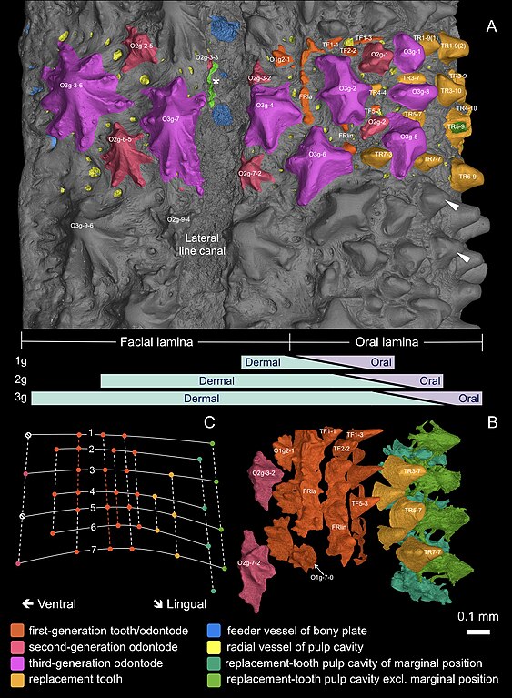
English: 3D external view of the scanned area. For three-dimensional curvature, see Figure 3. (A) External morphology with individually modeled structures highlighted in color. White arrowheads point to ornament-like teeth with side-cusps in the uncolored area. Bars in lavender and mint green indicate the putative gradient of the oral and dermal signal spheres, and the shift of the oral-dermal boundary during deposition of the first-generation (1g), second-generation (2g) and third-generation (3g) odontodes. (B) Overgrowing odontodes and bone matrix rendered invisible to show a consistent alternate pattern between replacement teeth, first-generation teeth and dermal odontodes dorsal to the lateral line canal. The replacement teeth at the inserted positions are not shown. Because the lingual rows of first-generation teeth are obscured by the labial rows of replacement teeth in this view, these rows are not shown (for all tooth rows, see Figure 5B). For optimal visibility, the most lingual rows of replacement teeth are represented by their pulp cavities. (C) Diagram of the alternate organization based on B. Solid lines, transverse files; numbers of files mark the putative level of the ossification center. Dashed lines, longitudinal rows; colored parts of the dashed lines indicate the range of the founder ridges. Dots denote positions of the structures in the particular colors. Note, the second-generation odontodes situated along the lingual border of the lateral line canal (O2g-3–2, O2g-7–2) have their labial ridgelets truncated, in order not to intrude on the canal sulcus. The alternate positions that are supposed to form the next row are suppressed (null signs), but the same alternate pattern develops properly on the other side of the canal (see Figure 4—figure supplement 1B). The dorsal edge of the odontodes at the ventral border of the canal is resorbed, leaving their pulp cavities open to the canal (asterisk; see Figure 3, O2g-3–3; Figure 4—figure supplement 1B, OP3-3, OP7-3; Figure 4—figure supplement 4). The ridgelets of the third-generation odontodes at both borders of the canal are also compressed (O3g-4, O3g-7). |
| Date | |
| Source | https://doi.org/10.7554/eLife.60985 |
| Author | Donglei Chen, Henning Blom, Sophie Sanchez, Paul Tafforeau, Tiiu Märss & Per E. Ahlberg |
Licensing
[edit]
This file is licensed under the Creative Commons Attribution 4.0 International license.
- You are free:
- to share – to copy, distribute and transmit the work
- to remix – to adapt the work
- Under the following conditions:
- attribution – You must give appropriate credit, provide a link to the license, and indicate if changes were made. You may do so in any reasonable manner, but not in any way that suggests the licensor endorses you or your use.
File history
Click on a date/time to view the file as it appeared at that time.
| Date/Time | Thumbnail | Dimensions | User | Comment | |
|---|---|---|---|---|---|
| current | 14:49, 13 August 2024 | | 1,654 × 2,254 (730 KB) | Trilletrollet (talk | contribs) | Uploaded a work by Donglei Chen, Henning Blom, Sophie Sanchez, Paul Tafforeau, Tiiu Märss & Per E. Ahlberg from https://doi.org/10.7554/eLife.60985 with UploadWizard |
You cannot overwrite this file.
File usage on Commons
There are no pages that use this file.