File:Liver and diaphragm covered in pedunculated growths Wellcome L0061574.jpg

Original file (4,542 × 6,330 pixels, file size: 7.5 MB, MIME type: image/jpeg)
Captions
Captions
Summary
[edit]
| Liver and diaphragm covered in pedunculated growths | |||
|---|---|---|---|
| Title |
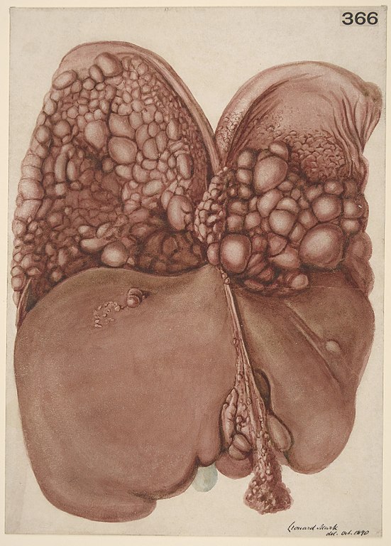
Liver and diaphragm covered in pedunculated growths |
||
| Description |
Watercolour drawing of the liver and of the under surface of the diaphragm. The latter is almost covered by a number of rounded pedunculated growths, varying in size from that of a walnut to that of a pea. There are also a few on the liver, and several on the peritoneal surface of the falciform ligament of the liver. Other parts of the peritoneum, especially that covering the uterus and ovaries, were thickly studded with similar growths. Under the microscope these were found to be composed of encephaloid carcinoma, the glandular cells being large and spheroidal in shape. The seat of the primary disease was not determined. Medical Photographic Library |
||
| Credit line |
|
||
| References |
|
||
| Source/Photographer |
https://wellcomeimages.org/indexplus/obf_images/af/e8/2bfb1e91b9c23f29c7c14fb6c5df.jpg
|
||
Licensing
[edit]
- You are free:
- to share – to copy, distribute and transmit the work
- to remix – to adapt the work
- Under the following conditions:
- attribution – You must give appropriate credit, provide a link to the license, and indicate if changes were made. You may do so in any reasonable manner, but not in any way that suggests the licensor endorses you or your use.
File history
Click on a date/time to view the file as it appeared at that time.
| Date/Time | Thumbnail | Dimensions | User | Comment | |
|---|---|---|---|---|---|
| current | 00:40, 18 October 2014 | | 4,542 × 6,330 (7.5 MB) | Fæ (talk | contribs) | =={{int:filedesc}}== {{Artwork |artist = |author = |title = Liver and diaphragm covered in pedunculated growths |description = Watercolour drawing of the liver and of the under surface of the diaphragm. The l... |
You cannot overwrite this file.
File usage on Commons
The following page uses this file: