File:Kidney tubules.png
Jump to navigation
Jump to search
No higher resolution available.
Kidney_tubules.png (256 × 256 pixels, file size: 118 KB, MIME type: image/png)
Captions
Captions
Add a one-line explanation of what this file represents
Summary
[edit]
| Description |
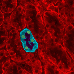
English: Microscopy of kidney tissue showing tubules. One tubule is highlighted to show epithelial cells (blue), cell nuclei (green) and the tubule lumen (dark center). Source: my personal image. The copyright to this image is retained by John Schmidt (JWSchmidt). Permission is granted to copy, distribute and/or modify this image under the terms of the Wikipedia GFDL, as indicated in the fine print at the bottom of this page. If you do not want to use this image under the terms of the GFDL, you can alternatively use it under the terms of the cc-by-nc-sa license.
Türkçe: Böbrek dokusu, tübüller gözükmekte. Tübüllerden biri epitel hücreleri (mavi), hücre çekirdeklerini (yeşil) ve tübül deliğini (koyu merkez/orta) göstermek için ışıklandırılmıştır. |
| Date | 2 April 2004 (original upload date) |
| Source | Transferred from en.wikipedia |
| Author | Original uploader was JWSchmidt at en.wikipedia |
| Permission (Reusing this file) |
GFDL-SELF-WITH-DISCLAIMERS; Released under the GNU Free Documentation License. |
Licensing
[edit]

|
Permission is granted to copy, distribute and/or modify this document under the terms of the GNU Free Documentation License, Version 1.2 or any later version published by the Free Software Foundation; with no Invariant Sections, no Front-Cover Texts, and no Back-Cover Texts. A copy of the license is included in the section entitled GNU Free Documentation License. |
| This file is licensed under the Creative Commons Attribution-Share Alike 3.0 Unported license. | ||
| ||
| This licensing tag was added to this file as part of the GFDL licensing update. |
I, the copyright holder of this work, hereby publish it under the following license:

|
Permission is granted to copy, distribute and/or modify this document under the terms of the GNU Free Documentation License, Version 1.2 or any later version published by the Free Software Foundation; with no Invariant Sections, no Front-Cover Texts, and no Back-Cover Texts. A copy of the license is included in the section entitled GNU Free Documentation License. Subject to disclaimers. |
Original upload log
[edit]
The original description page was here. All following user names refer to en.wikipedia.
- 2004-04-02 19:29 JWSchmidt 256×256×8 (121209 bytes) Microscopy of kidney tissue showing tubules.
File history
Click on a date/time to view the file as it appeared at that time.
| Date/Time | Thumbnail | Dimensions | User | Comment | |
|---|---|---|---|---|---|
| current | 15:10, 8 January 2008 | | 256 × 256 (118 KB) | Maderibeyza (talk | contribs) | {{Information |Description={{en|Microscopy of kidney tissue showing tubules. One tubule is highlighted to show epithelial cells (blue), cell nuclei (green) and the tubule lumen (dark center). Source: my personal image. The copyright to this image is retai |
You cannot overwrite this file.
File usage on Commons
The following page uses this file:
File usage on other wikis
The following other wikis use this file:
- Usage on ar.wikipedia.org
- Usage on bs.wikipedia.org
- Usage on en.wikipedia.org
- Usage on fa.wikipedia.org
- Usage on id.wikipedia.org
- Usage on sr.wikipedia.org
- Usage on ur.wikipedia.org
- Usage on zh.wikipedia.org