File:KidneyAndNephron-v4 Antares42.svg
Jump to navigation
Jump to search

Size of this PNG preview of this SVG file: 585 × 349 pixels. Other resolutions: 320 × 191 pixels | 640 × 382 pixels | 1,024 × 611 pixels | 1,280 × 764 pixels | 2,560 × 1,527 pixels.
Original file (SVG file, nominally 585 × 349 pixels, file size: 198 KB)
Captions
Captions
Add a one-line explanation of what this file represents
Summary
[edit]| Description |
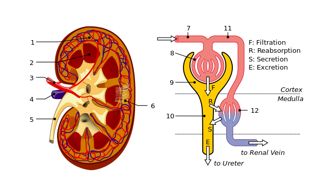
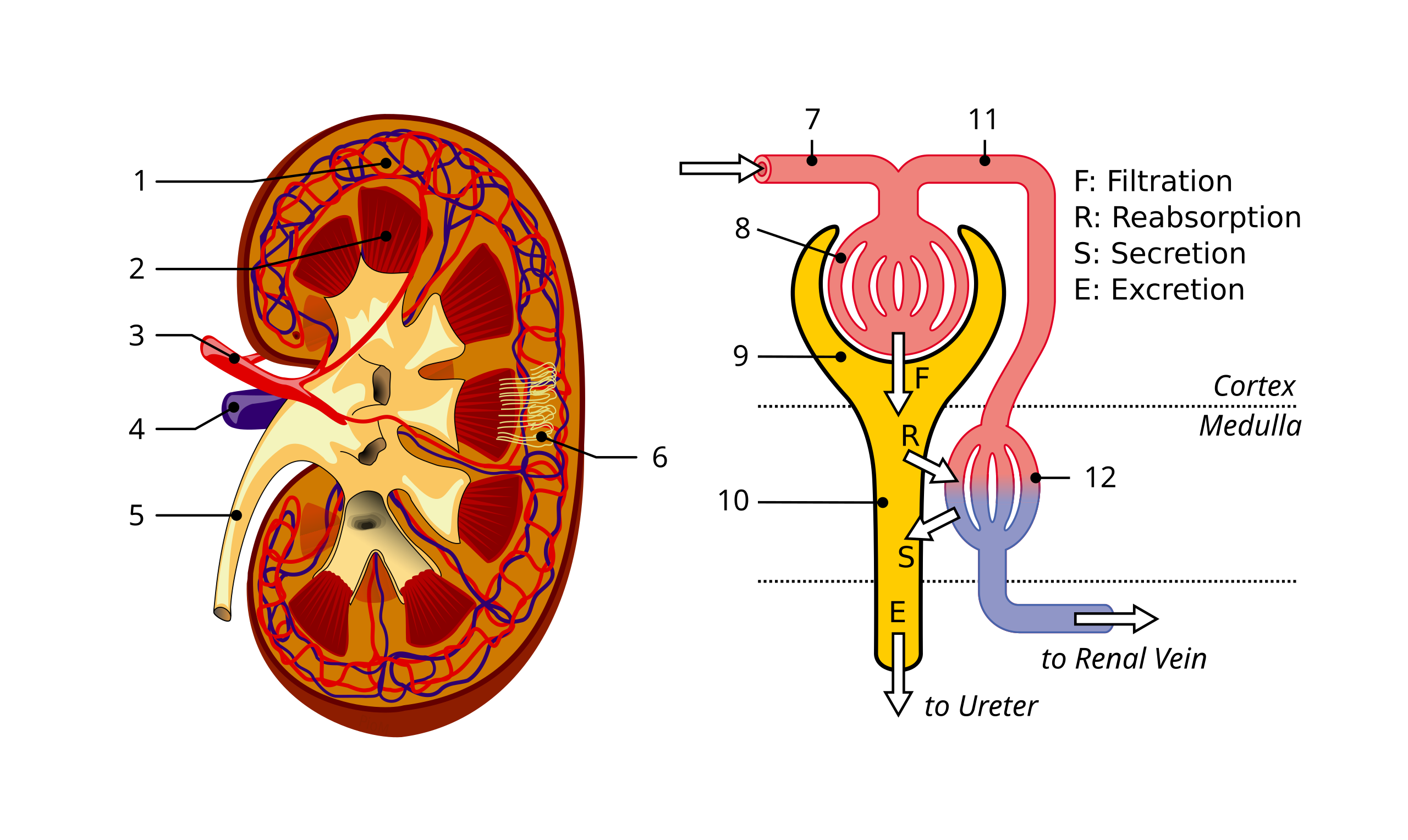
English: Schematics of kidney and nephron. 1: Renal cortex. 2: Medulla. 3: Renal artery. 4: Renal vein. 5: Ureter. 6: Nephrons. 7: Afferent arteriole. 8: Glomerulus. 9: Bowman’s capsule. 10: Tubuli and loop of Henle. 11: Efferent arteriole. 12: Peritubular capillaries. Français : Schémas du rein et d'un néphron. 1: Cortex rénal. 2: médullaire rénale. 3: Artère rénale. 4: Veine rénale. 5: Uretère. 6: Néphrons. 7: Artériole afférente . 8: Glomérule. 9: Capsule de Bowman. 10: Système tubulaire et anse de Henlé. 11: Artériole efférente. 12: Capillaires péritubulaires. |
| Date | (UTC) |
| Source |
This file was derived from: |
| Author |
|
| Other versions |
 |


| This is a retouched picture, which means that it has been digitally altered from its original version. Modifications: Merged kidney and nephron, simplified some of the legend. The original can be viewed here: KidneyStructures PioM.svg:
|
Licensing
[edit]
This file is licensed under the Creative Commons Attribution-Share Alike 3.0 Unported license.
- You are free:
- to share – to copy, distribute and transmit the work
- to remix – to adapt the work
- Under the following conditions:
- attribution – You must give appropriate credit, provide a link to the license, and indicate if changes were made. You may do so in any reasonable manner, but not in any way that suggests the licensor endorses you or your use.
- share alike – If you remix, transform, or build upon the material, you must distribute your contributions under the same or compatible license as the original.
Original upload log
[edit]
This image is a derivative work of the following images:
- File:Physiology_of_Nephron.svg licensed with Cc-by-3.0
- 2010-03-06T15:58:24Z Madhero88 1046x1221 (50398 Bytes) {{Information |Description={{en|1=[[:en:renal physiology|Physiology of Nephron]]}} |Source={{own}} |Author=[[User:Madhero88|Madhero88]] |Date= |Permission= |other_versions= }} [[Category:Nephrology]]
- File:KidneyStructures_PioM.svg licensed with Cc-by-sa-3.0-migrated, GFDL
- 2008-08-01T21:58:07Z Piom 2600x2640 (129606 Bytes) {{Information |Description= {{en| Structures of the kidney: #1.Renal pyramid #2.Interlobar artery #3.Renal artery #4.Renal vein #5.Renal hylum #6.Renal pelvis #7.Ureter #8.Minor calyx #9.Renal capsule #10.Inferior renal capsu
Uploaded with some help from derivativeFX

|
This SVG file contains embedded text that can be translated into your language, using any capable SVG editor, text editor or the SVG Translate tool. For more information see: About translating SVG files. |
File history
Click on a date/time to view the file as it appeared at that time.
| Date/Time | Thumbnail | Dimensions | User | Comment | |
|---|---|---|---|---|---|
| current | 18:02, 16 September 2013 | | 585 × 349 (198 KB) | Antares42 (talk | contribs) | == {{int:filedesc}} == {{Information |Description={{en| Schematics of kidney and nephron. 1: Renal cortex. 2: Medulla. 3: Renal artery. 4: Renal vein. 5: Ureter. 6: Nephrons. 7: Afferent arteriole. 8: Glomerulus. 9: Bowman’s capsule. 10: Tubuli and l... |
You cannot overwrite this file.
File usage on Commons
There are no pages that use this file.
File usage on other wikis
The following other wikis use this file:
- Usage on ru.wikipedia.org