File:Josef Reischig.jpg
Jump to navigation
Jump to search

Size of this preview: 492 × 599 pixels. Other resolutions: 197 × 240 pixels | 394 × 480 pixels | 630 × 768 pixels | 1,063 × 1,295 pixels.
Original file (1,063 × 1,295 pixels, file size: 369 KB, MIME type: image/jpeg)
Captions
Captions
Add a one-line explanation of what this file represents
Summary
[edit]
| Description |
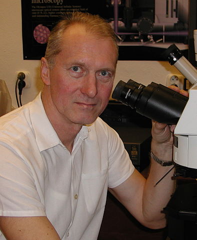
English: Josef Reischig on his work place - Department of Biology, Faculty of Medicine in Plzen, Charles University Čeština: Josef Reischig na svém pracovišti - Ústavu biologie Lékařské fakulty Univerzity Karlovy v Plzni |
| Date | |
| Source | Own work |
| Author | Petr Reischig |
| Permission (Reusing this file) |
Licensing
[edit]
I, the copyright holder of this work, hereby publish it under the following license:
This file is licensed under the Creative Commons Attribution-Share Alike 3.0 Unported license.
- You are free:
- to share – to copy, distribute and transmit the work
- to remix – to adapt the work
- Under the following conditions:
- attribution – You must give appropriate credit, provide a link to the license, and indicate if changes were made. You may do so in any reasonable manner, but not in any way that suggests the licensor endorses you or your use.
- share alike – If you remix, transform, or build upon the material, you must distribute your contributions under the same or compatible license as the original.
File history
Click on a date/time to view the file as it appeared at that time.
| Date/Time | Thumbnail | Dimensions | User | Comment | |
|---|---|---|---|---|---|
| current | 12:59, 7 November 2011 | | 1,063 × 1,295 (369 KB) | Petr Reischig (talk | contribs) |
You cannot overwrite this file.
File usage on Commons
More than 100 pages use this file. The following list shows the first 100 pages that use this file only. A full list is available.
- File:ADRENA 1 final.jpg
- File:Aspergillus fumigatus (257 15).jpg
- File:Bacteria (248 28) Airborne microbes.jpg
- File:Bacteria (251 31) Airborne microbes.jpg
- File:Bacteria (259 04) Airborne microbes.jpg
- File:Bacteria (259 05).jpg
- File:Bacteria (26 2 27) Airborne microbes.jpg
- File:Basidiobolus (257 20).jpg
- File:Candida albicans (248 35).jpg
- File:Candida albicans (251 12).jpg
- File:Candida albicans (257 12).jpg
- File:Cat fetus (247 30A) Cat fetus section.jpg
- File:Cat fetus (247 31A) Cat fetus section.jpg
- File:Cat fetus (26 2 03) Cat embryo longitudal section.jpg
- File:Cefftriaxon antibiotic.jpg
- File:Chromatin fibers (261 19) Mitosis; Xenopus egg.jpg
- File:Citric acid crystals.jpg
- File:Corynebacterium pseudodiphteriae (257 29).jpg
- File:Cross-cut bone.jpg
- File:Crystals of IgG antibodies.jpg
- File:Crystals of IgG antibodies 02.jpg
- File:Crystals of cytostatics Refador.jpg
- File:Crystals of vitamin B6.jpg
- File:Crystals of vitamin C.jpg
- File:Cyanobacteria (248 07) Mixture; native preparation; green filter.jpg
- File:Cyanobacteria (248 08) Mixture; native preparation; green filter.jpg
- File:Cyanobacteria (250 22) Native preparation.jpg
- File:Cyanobacteria (26 2 96) Native preparation.jpg
- File:Dopamin.jpg
- File:Duodenum (254 14).jpg
- File:Duodenum (26 2 08) Cross-section.jpg
- File:Escherichia coli (257 06) Gramnegative rods.jpg
- File:Escherichia coli (259 02) Gramnegative rods.jpg
- File:Escherichia coli (259 03) Gramnegative rods.jpg
- File:Etoposide 01.jpg
- File:Eubacteria (257 04) Various eubacteria.jpg
- File:Eubacteria (259 00F) Micrococcus luteus bacteria.jpg
- File:Eubacteria (259 01) Micrococcus luteus bacteria.jpg
- File:Eubacteria (259 10) Bacillus subtilis bacteria.jpg
- File:Eubacteria (259 11) Bacillus subtilis bacteria.jpg
- File:Eubacteria (259 12) Various eubacteria.jpg
- File:Eubacteria (259 13) Various eubacteria.jpg
- File:Eubacteria (263 09) Yoghurt bacteria.jpg
- File:Eubacteria (263 10) Bacterial capsule.jpg
- File:Eubacteria (263 11) Various eubacteria, spores stained green.jpg
- File:Eubacteria (26 2 26) Bacterial capsule.jpg
- File:Eubacteria (26 2 87) Bacillus subtilis; spores stained green.jpg
- File:Euglena sp (26 2 95).jpg
- File:Euglena sp (947 12).jpg
- File:Flea (251 01) Aphaniptera; total preparation.jpg
- File:Foraminifera (251 02) Mediterranean Sea.jpg
- File:Foraminifera (265 36) Various.jpg
- File:Glucose crystal.jpg
- File:Mabthera crystals.jpg
- File:Meiosis (248 22).jpg
- File:Meiosis (248 23).jpg
- File:Meiosis (254 30).jpg
- File:Meiosis (254 33).jpg
- File:Meiosis (261 23).jpg
- File:Meiosis (261 24).jpg
- File:Meiosis (261 25).jpg
- File:Meiosis (261 26).jpg
- File:Meiosis (261 27).jpg
- File:Meiosis (261 28).jpg
- File:Meiosis (261 29).jpg
- File:Meiosis (261 30).jpg
- File:Meiosis (261 32).jpg
- File:Meiosis (261 33).jpg
- File:Meiosis (261 34).jpg
- File:Meiosis (261 35).jpg
- File:Meiosis (261 36)- Dividing pollen mother cells M I, A I, T I (metaphase I, anaphase I, telophase I) - Lilium plant.jpg
- File:Meiosis (263 01).jpg
- File:Meiosis (263 02).jpg
- File:Meiosis (263 03).jpg
- File:Meiosis (263 04).jpg
- File:Meiosis (263 05).jpg
- File:Meiosis (263 07).jpg
- File:Meiosis (263 08).jpg
- File:Metaphase (261 04) Pressed; meristem of root; onion.jpg
- File:Metaphase (261 05) Pressed; meristem of root; onion.jpg
- File:Metaphase (261 06) Pressed; meristem of root; onion.jpg
- File:Metaphase (261 21) Pressed; meristem of root; onion.jpg
- File:Metaphase (261 22) Pressed; meristem of root; onion.jpg
- File:Plant epidermis (248 04) Epidermis of onion bulb; dry.jpg
- File:Plant epidermis (255 19) Epidermis of onion bulb ; wet.jpg
- File:Plant epidermis (255 20) Epidermis of onion bulb; dry.jpg
- File:Plant leaf epidermis (248 34) Tulip leaf epidermis.jpg
- File:Plant leaf epidermis (251 15) Upper epidermis of lime tree (Tilia).jpg
- File:Plant leaf epidermis (251 16) Lower epidermis of lime tree (Tilia).jpg
- File:Plant leaf epidermis (255 16) Tulip leaf epidermis.jpg
- File:Plant leaf epidermis (255 17) Lower epidermis of lime tree (Tilia).jpg
- File:Plant leaf epidermis (255 18) Upper epidermis of lime tree (Tilia).jpg
- File:Rat fetus (263 24A) Rat; section.jpg
- File:Snowflake crystal.jpg
- File:Thymus (265 25) Mammal.jpg
- File:Vessels (248 18) Cross-section.jpg
- Template:Josef Reischig
- Template:Josef Reischig/cs
- Template:Josef Reischig/en
- Template:Josef Reischig/layout
.jpg)
_Airborne_microbes.jpg)
_Airborne_microbes.jpg)
_Airborne_microbes.jpg)
.jpg)
_Airborne_microbes.jpg)
.jpg)
.jpg)
.jpg)
.jpg)
_Cat_fetus_section.jpg)
_Cat_fetus_section.jpg)
_Cat_embryo_longitudal_section.jpg)
_Mitosis;_Xenopus_egg.jpg)
.jpg)
_Mixture;_native_preparation;_green_filter.jpg)
_Mixture;_native_preparation;_green_filter.jpg)
_Native_preparation.jpg)
_Native_preparation.jpg)
.jpg)
_Cross-section.jpg)
_Gramnegative_rods.jpg)
_Gramnegative_rods.jpg)
_Gramnegative_rods.jpg)
_Various_eubacteria.jpg)
_Micrococcus_luteus_bacteria.jpg)
_Micrococcus_luteus_bacteria.jpg)
_Bacillus_subtilis_bacteria.jpg)
_Bacillus_subtilis_bacteria.jpg)
_Various_eubacteria.jpg)
_Various_eubacteria.jpg)
_Yoghurt_bacteria.jpg)
_Bacterial_capsule.jpg)
_Various_eubacteria,_spores_stained_green.jpg)
_Bacterial_capsule.jpg)
_Bacillus_subtilis;_spores_stained_green.jpg)
.jpg)
.jpg)
_Aphaniptera;_total_preparation.jpg)
_Mediterranean_Sea.jpg)
_Various.jpg)
.jpg)
.jpg)
.jpg)
.jpg)
.jpg)
.jpg)
.jpg)
.jpg)
.jpg)
.jpg)
.jpg)
.jpg)
.jpg)
.jpg)
.jpg)
.jpg)
-_Dividing_pollen_mother_cells_M_I,_A_I,_T_I_(metaphase_I,_anaphase_I,_telophase_I)_-_Lilium_plant.jpg)
.jpg)
.jpg)
.jpg)
.jpg)
.jpg)
.jpg)
.jpg)
_Pressed;_meristem_of_root;_onion.jpg)
_Pressed;_meristem_of_root;_onion.jpg)
_Pressed;_meristem_of_root;_onion.jpg)
_Pressed;_meristem_of_root;_onion.jpg)
_Pressed;_meristem_of_root;_onion.jpg)
_Epidermis_of_onion_bulb;_dry.jpg)
_Epidermis_of_onion_bulb_;_wet.jpg)
_Epidermis_of_onion_bulb;_dry.jpg)
_Tulip_leaf_epidermis.jpg)
_Upper_epidermis_of_lime_tree_(Tilia).jpg)
_Lower_epidermis_of_lime_tree_(Tilia).jpg)
_Tulip_leaf_epidermis.jpg)
_Lower_epidermis_of_lime_tree_(Tilia).jpg)
_Upper_epidermis_of_lime_tree_(Tilia).jpg)
_Rat;_section.jpg)
_Mammal.jpg)
_Cross-section.jpg)
View more links to this file.
File usage on other wikis
The following other wikis use this file:
- Usage on cs.wikipedia.org
- Usage on www.wikidata.org