File:Holothuria tubulosa development embryo two stages.jpg
Original file (1,079 × 688 pixels, file size: 735 KB, MIME type: image/jpeg)
Captions
Captions
Summary
[edit]
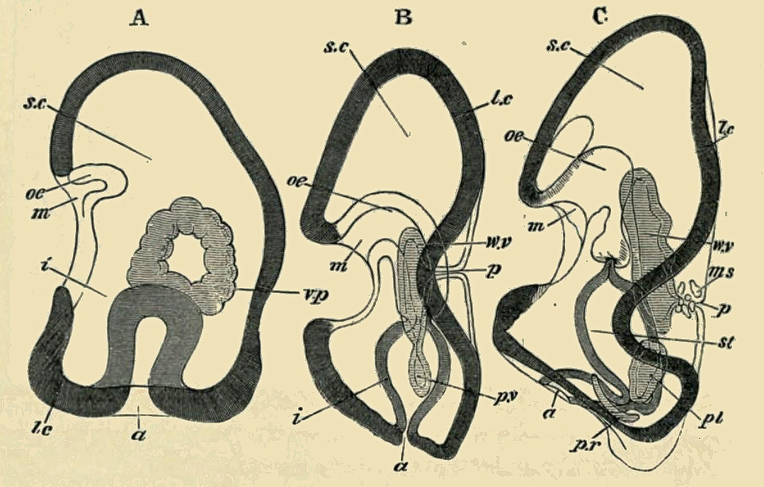
| Description | FIG. 248. THREE STAGES IN THE DEVELOPMENT OF HOLOTHURIA TUBULOSA VIEWED FROM THE SIDE IN OPTICAL SECTION. (After Selenka.) m. mouth; oe. oesophagus; st. stomach; i. intestine; a. anus; l.c. longitudinal ciliated band; s.c. segmentation cavity; v.p. vaso-peritoneal vesicle; p.v. peritoneal vesicle; p.r. right peritoneal vesicle ; pl. left peritoneal vesicle ; w.v. water- vascular vesicle ; p. dorsal pore of water- vascular system ; ms. muscle cells. |
| Date | |
| Source |
https://archive.org/details/theworks02balfuoft/page/546/mode/2up?view=theater&q=blastoderm THE WORKS OF FRANCIS MAITLAND BALFOUR VOL. III A TREATISE ON COMPARATIVE EMBRYOLOGY. |
| Author | M. FOSTER, F.R.S., ADAM SEDGWICK, M.A., |
Licensing
[edit]
|
This work is in the public domain in its country of origin and other countries and areas where the copyright term is the author's life plus 70 years or fewer.
| |
| This file has been identified as being free of known restrictions under copyright law, including all related and neighboring rights. | |

|
This file, which was originally posted to an external website, has not yet been reviewed by an administrator or reviewer to confirm that the above license is valid. See Category:License review needed for further instructions.
|
File history
Click on a date/time to view the file as it appeared at that time.
| Date/Time | Thumbnail | Dimensions | User | Comment | |
|---|---|---|---|---|---|
| current | 22:13, 2 March 2024 | | 1,079 × 688 (735 KB) | Rasbak (talk | contribs) | {{Information |description=FIG. 248. THREE STAGES IN THE DEVELOPMENT OF HOLOTHURIA TUBULOSA VIEWED FROM THE SIDE IN OPTICAL SECTION. (After Selenka.) tn. mouth; oe. oesophagus; st. stomach; i. intestine; a. anus; l.c. longitudinal ciliated band; v.p. vaso-peritoneal vesicle; p.v. peritoneal vesicle; p.r. right peritoneal vesicle ; pl. left peritoneal vesicle ; w.v. water- vascular vesicle ; p. dorsal pore of water- vascular system ; ms. muscle cells. |source=https://archive.org/details/thewor... |
You cannot overwrite this file.
File usage on Commons
The following 2 pages use this file: