File:Histopathology of progressive atherosclerotic lesion with fibrous cap and necrotic core.jpg
Jump to navigation
Jump to search
Size of this preview: 800 × 304 pixels. Other resolutions: 320 × 122 pixels | 892 × 339 pixels.
Original file (892 × 339 pixels, file size: 150 KB, MIME type: image/jpeg)
Captions
Captions
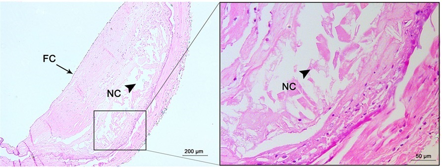
Histopathology of progressive atherosclerotic lesion with fibrous cap and necrotic core
Summary
[edit]| Description |
English: Histopathology of a progressive atherosclerotic lesion: Fibrous cap atheroma (B) has a well-formed necrotic core (NC) containing lipids with an overlying thick fibrous cap (FC). Hematoxylin and eosin (H&E) staining; original magnification: 5× for original images, 20× for inserted images. |
| Date | |
| Source |
(2017). "Histological Characteristics of Intracranial Atherosclerosis in a Chinese Population: A Postmortem Study". Frontiers in Neurology 8. DOI:10.3389/fneur.2017.00488. ISSN 1664-2295. - Attribution 4.0 International (CC BY 4.0) license |
| Author | Yang, Wen Jie; Fisher, Mark; Zheng, Lu; Niu, Chun Bo; Paganini-Hill, Annlia; Zhao, Hai Lu; Xu, Yun; Wong, Ka Sing; Ng, Ho Keung; Chen, Xiang Yan |
| Other versions |
|

Licensing
[edit]
This file is licensed under the Creative Commons Attribution 4.0 International license.
- You are free:
- to share – to copy, distribute and transmit the work
- to remix – to adapt the work
- Under the following conditions:
- attribution – You must give appropriate credit, provide a link to the license, and indicate if changes were made. You may do so in any reasonable manner, but not in any way that suggests the licensor endorses you or your use.
File history
Click on a date/time to view the file as it appeared at that time.
| Date/Time | Thumbnail | Dimensions | User | Comment | |
|---|---|---|---|---|---|
| current | 19:35, 29 December 2020 | 892 × 339 (150 KB) | Mikael Häggström (talk | contribs) | Uploaded a work by Yang, Wen Jie; Fisher, Mark; Zheng, Lu; Niu, Chun Bo; Paganini-Hill, Annlia; Zhao, Hai Lu; Xu, Yun; Wong, Ka Sing; Ng, Ho Keung; Chen, Xiang Yan from {{cite journal|last1=Yang|first1=Wen Jie|last2=Fisher|first2=Mark|last3=Zheng|first3=Lu|last4=Niu|first4=Chun Bo|last5=Paganini-Hill|first5=Annlia|last6=Zhao|first6=Hai Lu|last7=Xu|first7=Yun|last8=Wong|first8=Ka Sing|last9=Ng|first9=Ho Keung|last10=Chen|first10=Xiang Yan|title=Histological Characteristics of Intracranial Athe... |
You cannot overwrite this file.
File usage on Commons
The following 5 pages use this file:
- File:Histopathology of a fibrocalcific atheroma.jpg
- File:Histopathology of progressive atherosclerotic lesion with extracellular lipid.jpg
- File:Histopathology of progressive atherosclerotic lesion with fibrous cap and necrotic core.jpg
- File:Histopathology of progressive atherosclerotic lesions.jpg
- Template:Progressive atherosclerotic lesions - versions