File:Histopathology of nodular melanoma.jpg
Jump to navigation
Jump to search
Size of this preview: 800 × 298 pixels. Other resolutions: 320 × 119 pixels | 1,183 × 441 pixels.
Original file (1,183 × 441 pixels, file size: 212 KB, MIME type: image/jpeg)
Captions
Captions
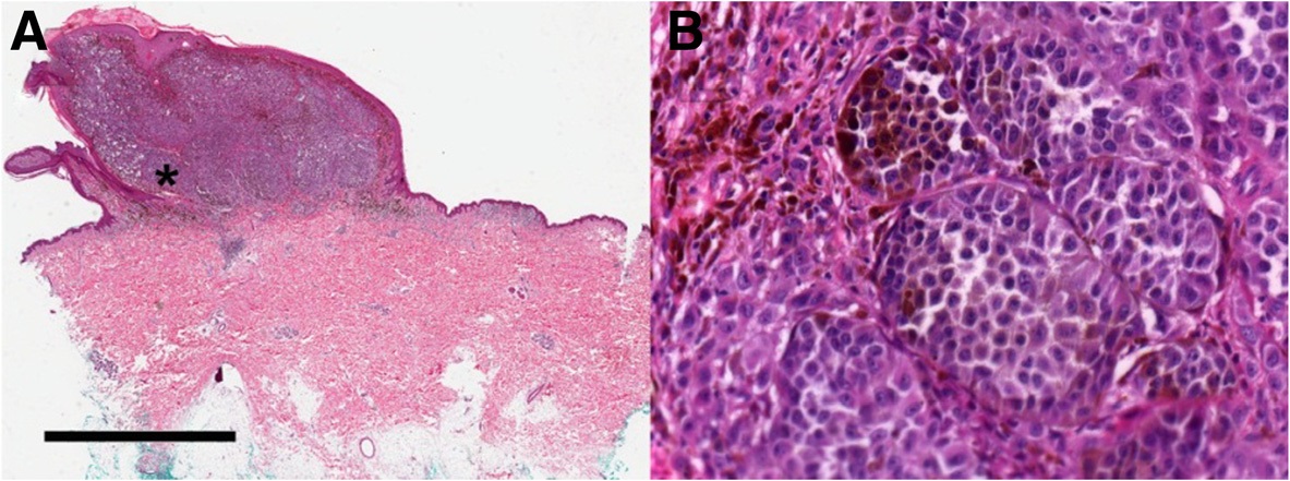
Photomicrograph of H&E stained primary nodular melanoma at (a) low and (b) high magnification showing nests of atypical melanocyte proliferations
Summary
[edit]
| Description |
English: Photomicrograph of H&E stained primary nodular melanoma at (a) low and (b) high magnification showing nests of atypical melanocyte proliferations. The scale bar represents 4 mm for panel A and 125 μM for panel B. |
| Date | |
| Source |
(2015). "Somatic and germline analyses of a long term melanoma survivor with a recurrent brain metastasis". BMC Cancer 15 (1). DOI:10.1186/s12885-015-1927-0. ISSN 1471-2407. - "This article is distributed under the terms of the Creative Commons Attribution 4.0 International License (https://creativecommons.org/licenses/by/4.0/)" |
| Author | Weiss, Sarah; Darvishian, Farbod; Tadepalli, Jyothi; Shapiro, Richard; Golfinos, John; Pavlick, Anna; Polsky, David; Kirchhoff, Tomas; Osman, Iman |
| Other versions |
 |
Licensing
[edit]
This file is licensed under the Creative Commons Attribution 4.0 International license.
- You are free:
- to share – to copy, distribute and transmit the work
- to remix – to adapt the work
- Under the following conditions:
- attribution – You must give appropriate credit, provide a link to the license, and indicate if changes were made. You may do so in any reasonable manner, but not in any way that suggests the licensor endorses you or your use.
File history
Click on a date/time to view the file as it appeared at that time.
| Date/Time | Thumbnail | Dimensions | User | Comment | |
|---|---|---|---|---|---|
| current | 16:01, 25 February 2020 | 1,183 × 441 (212 KB) | Mikael Häggström (talk | contribs) | User created page with UploadWizard |
You cannot overwrite this file.
File usage on Commons
The following 2 pages use this file:
File usage on other wikis
The following other wikis use this file:
- Usage on en.wikipedia.org
- Usage on eu.wikipedia.org