File:Histopathology of meningioma.png
Jump to navigation
Jump to search

Size of this preview: 800 × 598 pixels. Other resolutions: 320 × 239 pixels | 640 × 479 pixels | 1,024 × 766 pixels | 1,280 × 958 pixels | 2,048 × 1,532 pixels.
Original file (2,048 × 1,532 pixels, file size: 5.48 MB, MIME type: image/png)
Captions
Captions
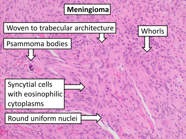
Histopathology of meningioma, H&E stain
Summary
[edit]
| Description |
English: Histopathology of a typical WHO grade 1 meningioma, H&E stain. It is of the meningothelial histologic type, which is the most common meningioma type. It shows its typical findings: - A woven architectural pattern - Psammoma bodies (spheroid calcifications) - Syncytial cells (having indistinct cell membranes) with eosinophilic (pink) cytoplasms - Round uniform nuclei - Whorls (concentric cell arrangements)
|
| Date | |
| Source | Own work |
| Author |
.jpg) - Reusing images - Conflicts of interest: None Consent note: Consent from the patient or patient's relatives is regarded as redundant, because of absence of identifiable features (List of HIPAA identifiers) in the media and case information (See also HIPAA case reports guidance). |
| Other versions |
 |
Licensing
[edit]
| This file is made available under the Creative Commons CC0 1.0 Universal Public Domain Dedication. | |
| The person who associated a work with this deed has dedicated the work to the public domain by waiving all of their rights to the work worldwide under copyright law, including all related and neighboring rights, to the extent allowed by law. You can copy, modify, distribute and perform the work, even for commercial purposes, all without asking permission.
|
File history
Click on a date/time to view the file as it appeared at that time.
| Date/Time | Thumbnail | Dimensions | User | Comment | |
|---|---|---|---|---|---|
| current | 17:17, 14 January 2024 | | 2,048 × 1,532 (5.48 MB) | Mikael Häggström (talk | contribs) | To trabecular |
| 19:30, 11 January 2024 |  | 2,048 × 1,532 (5.61 MB) | Mikael Häggström (talk | contribs) | Eosinophilic | |
| 19:25, 11 January 2024 |  | 2,048 × 1,532 (5.91 MB) | Mikael Häggström (talk | contribs) | Uploaded a work by {{Mikael Häggström|cat=Micrographs of the brain|consent=noid}} from {{Own}} with UploadWizard |
You cannot overwrite this file.
File usage on Commons
The following page uses this file:
File usage on other wikis
The following other wikis use this file:
- Usage on en.wikipedia.org