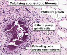
File:Histopathology of calcifying aponeurotic fibroma, original.png

Original file (785 × 647 pixels, file size: 1.07 MB, MIME type: image/png)
Captions
Captions
Summary
[edit]| DescriptionHistopathology of calcifying aponeurotic fibroma, original.png |
English: Histopathology of a calcifying aponeurotic fibroma from a finger, H&E stain. Detail from a whole slide scan. |
| Date | |
| Source | Case 15 - Calcifying aponeurotic fibroma. AceMyPath at Pathpresenter. Retrieved on 2023-07-10. |
| Author | Rajendra Singh, Kamran Mirza, Kurt Schaberg |
| Other versions |
 |
Licensing
[edit]| Public domainPublic domainfalsefalse |
| This file is in the public domain because it is a scan of a microscopy slide created in the United States and does not contain additional copyrightable graphics. See Meta:Wikilegal/Copyright of Medical Imaging for details.
|
||
| This file has been identified as being free of known restrictions under copyright law, including all related and neighboring rights. | ||
https://creativecommons.org/publicdomain/mark/1.0/PDMCreative Commons Public Domain Mark 1.0falsefalse
File history
Click on a date/time to view the file as it appeared at that time.
| Date/Time | Thumbnail | Dimensions | User | Comment | |
|---|---|---|---|---|---|
| current | 23:11, 10 July 2023 |  | 785 × 647 (1.07 MB) | Mikael Häggström (talk | contribs) | Uploaded own work with UploadWizard |
You cannot overwrite this file.
File usage on Commons
The following page uses this file:
Metadata
This file contains additional information such as Exif metadata which may have been added by the digital camera, scanner, or software program used to create or digitize it. If the file has been modified from its original state, some details such as the timestamp may not fully reflect those of the original file. The timestamp is only as accurate as the clock in the camera, and it may be completely wrong.
| Horizontal resolution | 37.79 dpc |
|---|---|
| Vertical resolution | 37.79 dpc |
