File:Hemienchytraeus wuhanensis (10.3897-zookeys.1015.59019) Figure 3.jpg
Jump to navigation
Jump to search

Size of this preview: 794 × 600 pixels. Other resolutions: 318 × 240 pixels | 636 × 480 pixels | 1,017 × 768 pixels | 1,280 × 967 pixels | 1,512 × 1,142 pixels.
Original file (1,512 × 1,142 pixels, file size: 2.57 MB, MIME type: image/jpeg)
Captions
Captions
Add a one-line explanation of what this file represents
Summary
[edit]_Figure_3.jpg&action=edit§ion=1)
| Description |
English: Figure 3.
|
| Date | |
| Source | https://doi.org/10.3897/zookeys.1015.59019 |
| Author | Chen J, Schmelz RM, Xie Z (2021) Description of Hemienchytraeus wuhanensis sp. nov. (Annelida, Clitellata, Enchytraeidae) from central China, with comments on species records of Hemienchytraeus from China. ZooKeys 1015: 87-97. |
| Permission (Reusing this file) |
This file is licensed under the Creative Commons Attribution 4.0 International license.
|
File history
Click on a date/time to view the file as it appeared at that time.
| Date/Time | Thumbnail | Dimensions | User | Comment | |
|---|---|---|---|---|---|
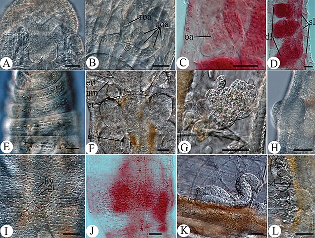
| current | 06:12, 25 September 2022 | | 1,512 × 1,142 (2.57 MB) | Christian Ferrer (talk | contribs) | {{Information | description = {{en|1=Figure 3. :Micrographs of ''Hemienchytraeus wuhanensis'' sp. nov. A, B, E–I, K, L in vivo C, D, J fixed A brain B dorsal view of oesophageal appendage C lateral view of oesophageal appendage D pharyngeal glands E epidermal gland cells in II–V ventrally F spermathecae and pharyngeal glands G nephridia in 7/8, anteseptale bottom-left H male glandular bulb, slightly everted I dorsal view of clitellum J ventral view of clitellum K sperm funnel L sperm duct a... |
You cannot overwrite this file.
File usage on Commons
There are no pages that use this file.
_Figure_3.jpg&oldid=720907412)