File:HIV-budding-BW-detail(2).jpg
Jump to navigation
Jump to search

Size of this preview: 760 × 600 pixels. Other resolutions: 304 × 240 pixels | 608 × 480 pixels | 973 × 768 pixels | 1,280 × 1,010 pixels | 2,560 × 2,020 pixels | 2,760 × 2,178 pixels.
Original file (2,760 × 2,178 pixels, file size: 943 KB, MIME type: image/jpeg)
Captions
Captions
Add a one-line explanation of what this file represents
Summary
[edit]| Description |
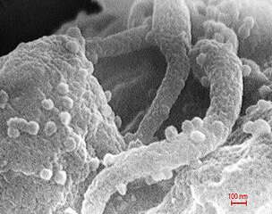
English: This scanning electron micrograph revealed the presence of the human immunodeficiency virus (HIV-1), (spherical in appearance), which had been co-cultivated with human lymphocytes. Note the lymphocyte in the lower left, and some of its extended pseudopodia. HIV-1 virions can be seen on the surface of this lymphocyte. See PHIL 1843 for another view of this electron microscopic scenario.
A retrovirus, the Human Immunodeficiency Virus (HIV) was identified in 1983 as the pathogen responsible for the Acquired Immunodeficiency Syndrome (AIDS). AIDS is characterized by changes in the population of T-cell lymphocytes that play a key role in the immune defense system. In the infected individual, the virus causes a depletion of T-cells, called “T-helper cells”, which leaves these patients susceptible to opportunistic infections, and certain malignancies. |
||
| Date | |||
| Source |
|
||
| Author |
|
||
| Permission (Reusing this file) |
PD-USGov-HHS-CDC English: None - This image is in the public domain and thus free of any copyright restrictions. As a matter of courtesy we request that the content provider be credited and notified in any public or private usage of this image. |
||
| Other versions |
|


.jpg)
.jpg&action=edit§ion=1)
Licensing
[edit].jpg&action=edit§ion=2)
This image is a work of the Centers for Disease Control and Prevention, part of the United States Department of Health and Human Services, taken or made as part of an employee's official duties. As a work of the U.S. federal government, the image is in the public domain.
|
File history
Click on a date/time to view the file as it appeared at that time.
| Date/Time | Thumbnail | Dimensions | User | Comment | |
|---|---|---|---|---|---|
| current | 08:08, 16 November 2008 | | 2,760 × 2,178 (943 KB) | Optigan13 (talk | contribs) | == Summary == {{Information |Description={{en|This scanning electron micrograph revealed the presence of the human immunodeficiency virus (HIV-1), (spherical in appearance), which had been co-cultivated with human lymphocytes. Note the lymphocyte in the l |
You cannot overwrite this file.
File usage on Commons
There are no pages that use this file.
File usage on other wikis
The following other wikis use this file:
- Usage on es.wikipedia.org
.jpg&oldid=859927088)