File:Gray881.png
Gray881.png (500 × 335 pixels, file size: 29 KB, MIME type: image/png)
Captions
Captions
Summary
[edit]| Description |
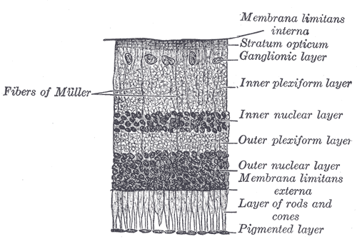
Original description from http://www.bartleby.com/107/225.html. The retina consists of an outer pigmented layer and an inner nervous stratum or retina proper. The pigmented layer consists of a single stratum of cells. When viewed from the outer surface these cells are smooth and hexagonal in shape; when seen in section each cell consists of an outer non-pigmented part containing a large oval nucleus and an inner pigmented portion which extends as a series of straight thread-like processes between the rods, this being especially the case when the eye is exposed to light. In the eyes of albinos the cells of this layer are destitute of pigment. Retina Proper —The nervous structures of the retina proper are supported by a series of nonnervous or sustentacular fibers, and, when examined microscopically by means of sections made perpendicularly to the surface of the retina, are found to consist of seven layers, named from within outward as follows:
|
||||||||||||||||||||
| Plate | 881 | ||||||||||||||||||||
| Date | before 1858 | ||||||||||||||||||||
| Source |
|
||||||||||||||||||||
| Author |
|
||||||||||||||||||||
| Other versions | Derivative works of this file: Gray881-ar.png | ||||||||||||||||||||
.jpg)
Book
[edit]| Henry Gray: Gray's Anatomy (20th edition)
|
|||||||||||||||||||||||
|---|---|---|---|---|---|---|---|---|---|---|---|---|---|---|---|---|---|---|---|---|---|---|---|
| Author |
|
-_Title_page.png) | |||||||||||||||||||||
| Editor |
Revised by Warren H. Lewis |
||||||||||||||||||||||
| Illustrator |
|
||||||||||||||||||||||
| Title | |||||||||||||||||||||||
| Edition |
20 |
||||||||||||||||||||||
| Publisher | |||||||||||||||||||||||
| Object type |
version, edition or translation |
||||||||||||||||||||||
| Page overview | list of all the plates | ||||||||||||||||||||||
| Language |
English |
||||||||||||||||||||||
| Publication date |
1918 |
||||||||||||||||||||||
| Place of publication |
Philadelphia / New York City |
||||||||||||||||||||||
| Source | Bartleby | ||||||||||||||||||||||

Licensing
[edit]
This image is in the public domain because it is a mere mechanical scan or photocopy of a public domain original, or – from the available evidence – is so similar to such a scan or photocopy that no copyright protection can be expected to arise. The original itself is in the public domain for the following reason:
This tag is designed for use where there may be a need to assert that any enhancements (eg brightness, contrast, colour-matching, sharpening) are in themselves insufficiently creative to generate a new copyright. It can be used where it is unknown whether any enhancements have been made, as well as when the enhancements are clear but insufficient. For known raw unenhanced scans you can use an appropriate {{PD-old}} tag instead. For usage, see Commons:When to use the PD-scan tag.  | ||||
File history
Click on a date/time to view the file as it appeared at that time.
| Date/Time | Thumbnail | Dimensions | User | Comment | |
|---|---|---|---|---|---|
| current | 21:19, 23 January 2007 | | 500 × 335 (29 KB) | Pngbot (talk | contribs) | optimized with optipng |
| 03:14, 11 February 2006 |  | 500 × 335 (50 KB) | Arcadian (talk | contribs) | {{Gray's Anatomy plate}} |
You cannot overwrite this file.
File usage on Commons
The following 3 pages use this file:
File usage on other wikis
The following other wikis use this file:
- Usage on ar.wikipedia.org
- Usage on bg.wikipedia.org
- Usage on bn.wikipedia.org
- Usage on bs.wikipedia.org
- Fotoosjetljive ganglijske ćelije
- Unutrašnja granična membrana
- Sloj nervnih vlakana
- Sloj ganglijskih ćelija
- Unutrašnji pleksiformni sloj
- Unutrašnji jezgarni sloj
- Vanjski pleksiformni sloj
- Vanjski jezgarni sloj
- Vanjska granična membrana
- Sloj štapića i čepića
- Pigmentni epitel mrežnjače
- Urođeno stacionarno noćno sljepilo
- Usage on ckb.wikipedia.org
- Usage on de.wikibooks.org
- Usage on en.wikipedia.org
- Retina
- Neuropil
- Inner plexiform layer
- Layer of rods and cones
- Retinal pigment epithelium
- Inner nuclear layer
- External limiting membrane
- Internal limiting membrane
- Outer plexiform layer
- Outer nuclear layer
- Retinal nerve fiber layer
- Ganglion cell layer
- Congenital stationary night blindness
- Retinal regeneration
- User:Was a bee/Gray
- Usage on fa.wikipedia.org
- Usage on he.wikipedia.org
- Usage on hr.wikipedia.org
- Usage on hy.wikipedia.org
- Usage on it.wikipedia.org
- Usage on ja.wikipedia.org
- Usage on kn.wikipedia.org
- Usage on ko.wikipedia.org
View more global usage of this file.