File:Gallus gallus domesticus (02).jpg
Original file (781 × 1,243 pixels, file size: 1.09 MB, MIME type: image/jpeg)
Captions
Captions
Summary
[edit].jpg&action=edit§ion=1)
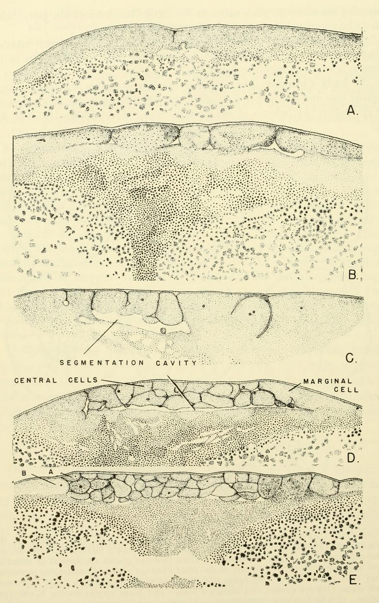
| Description | Fig. 156. Cleavage in chick blastoderm, sectional views. (After Patterson, '10.) (A) Median section through blastoderm approximately at right angles to furrow shown in fig. 155 A. (B) Section through blastoderm of about eight-cell stage. (C) Section through blastoderm, showing 32 cells, also showing horizontal cytoplasmic cleft (segmentation cavity). (D) Median section through blastoderm similar to that shown in fig. 155E. (E) Median section through blastoderm similar to that of fig. 155G. (F, G) Diagrammatic views of developing avian blastoderms. (F) Diagrammatic section and surface view of chick blastoderm shown in fig. 155G and fig. 156E. (G) Section of chick blastoderm about time that egg is laid, depicting the primary blastocoel below the blastoderm and syncytial tissue at the margins. Observe that the syncytial tissue serves to implant the blastoderm upon the yolk substance. |
| Date | |
| Source | https://archive.org/details/comparativeembry00nels/page/314/mode/1up?view=theater&q=BLASTULATION+ Comparative embryology of the vertebrates; with 2057 drawings and photos. grouped as 380 illustrations. |
| Author | Nelsen, Olin E. |
Licensing
[edit].jpg&action=edit§ion=2)
|
This work is in the public domain in its country of origin and other countries and areas where the copyright term is the author's life plus 70 years or fewer.
| |
| This file has been identified as being free of known restrictions under copyright law, including all related and neighboring rights. | |

|
This file, which was originally posted to an external website, has not yet been reviewed by an administrator or reviewer to confirm that the above license is valid. See Category:License review needed for further instructions.
|
File history
Click on a date/time to view the file as it appeared at that time.
| Date/Time | Thumbnail | Dimensions | User | Comment | |
|---|---|---|---|---|---|
| current | 11:02, 11 March 2024 | | 781 × 1,243 (1.09 MB) | Rasbak (talk | contribs) | {{Information |description=Fig. 156. Cleavage in chick blastoderm, sectional views. (After Patterson, '10.) (A) Median section through blastoderm approximately at right angles to furrow shown in fig. 155 A. (B) Section through blastoderm of about eight-cell stage. (C) Section through blastoderm, showing 32 cells, also showing horizontal cytoplasmic cleft (segmentation cavity). (D) Median section through blastoderm similar to that shown in fig. 155E. (E) Median section through blastoderm simil... |
You cannot overwrite this file.
File usage on Commons
There are no pages that use this file.
.jpg&oldid=859744297)