File:Fletcher Oxycephaly 6.jpg
Original file (892 × 1,094 pixels, file size: 91 KB, MIME type: image/jpeg)
Captions
Captions
Summary
[edit]
| Description |
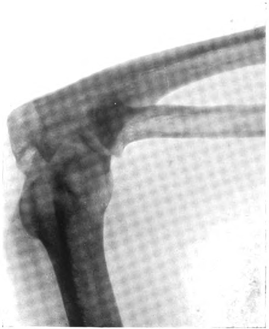
English: Case 1. A. R., male, 23, storekeeper : photographs (Plate 30), skiagram of
skul? (Plate 31). Did not know at what age the exophthalmos or head deformity begen. Eyesight defective as long as he could remember, and always worse in left eye. Never went to school, owing to bad eyesight, but taught himself to read and write with his father's help. Did not suffer from headache and had never hali fits. His mother (whom I have seen) had exophthalmos and malformation of the superior maxilla, but the vault of the cranium was not definitely oxy- cophalic. Parents English. Head typically oxycephalic (photographs, Plate 30). The bregma was promi. nent. Extreme proptosis; the right eye was more prominent than the left and occasionally became dislocated forwards in front of the eyelids. Divergent squint and nystagmus, could distinguish only moving objects with left eye, can read print with right eye. Mr. Holmes Spicer reports : Right pupil unusually small, but reacts normally. Left pupil larger than right and insensitive to light. Both optic disks very white; the right disk has ill-defined edges and a definite white fibrosis-looking prolongation up and inwards suggesting a previous neuritis; the lamina cribrosa also is not visible, but the vessels are quite clear. The left disk is quite clear and the vessels well defined—the lamina cribrosa is not seen. The condition of the disks is quite compatible with previous neuritis. Right visual field much reduced.' The nose was strongly deflected to the right; this was stated to be the result of an injury. The superior maxilla was very deformed, it was shortened from before backwards. The hard palate formed a narrow, very pointed arch. The second and third molars were absent in the upper jaw and could be made out in the skiagram lying above in what appeared to be the rudimentary antra. Lower jaw underhung, teeth normal in number. Sense of smell lost. Hearing and taste normal. Elbows could not be fully extended (Plate 30, Fig. 3, and Plate 31, Figs. 5 and 6). Internal condyles unduly prominent, some creaking in the joints : the head of both radius and ulna appears thickened. He could not raise the arms to the horizontal; obvious creaking in both shoulder-joints, but no change found in the bones. No other malformations present. Skiagram (Plate 31, Fig. 4) of the skull shows very distinctly the bulging and thinning of the bone in the bregmatic region (?) and the digital markings over the whole of the vault. The most striking abnormality is the altered shape and depth of the middle fossa, and the pushing forward of the posterior wall of the orbit. The sella turcica is pushed backwards and is deepened (cf. skiagram of normal adult skull, Plate 38, Fig. 23). Dr. R. J. Gladstone very kindly took the following measurements of this case two years ago : Height, 5 ft. 22 in. (1522 mm.). Circumference of head, 506 mm. Longitudinal arc, 345 mm. Transverse arc, 352 mm. Diameters of head: L, 179 mm. B. 138 mm. H. 143 mm. Mi. F. 106 mm. Index of size' of head, 3532. (The average 'index of size of males, 5 ft. 3 in. height, is 3850.) Cephalic breadth index, 76. Cephalic height index, 79.8. Horizontal arc, measured from the centre of the external auditory meatus round the sub-nasal point to the centre of the meatus of the opposite side = 231 min. (Average about 255 mm.) Average diameters of head in fifty male subjects, aged 20 to 46, measured in the post-mortem room, Middlesex Hospital :- Length: glabella to occipital point . . 190.8 mm. Breadth : greatest transverse diameter . . 149.5 mm. Height: bi-auricular line to vertex . 134.8 mm. IQ. J. M., April, 1911.) D d There is thus considerable diminution in the length and breadth, and increase in height. Normal Case A. R. Average cephalic breadth index . . 77.6 76.0 » » height , . . . 70.0 79.8 |
| Date | between 1910 and 1911 |
| Source | "On Oxycephaly", Th Quarterly Journal of Medicine, Volume IV |
| Author | H. Morley Fletcher |
Licensing
[edit]
|
This work is in the public domain in its country of origin and other countries and areas where the copyright term is the author's life plus 70 years or fewer. This work is in the public domain in the United States because it was published (or registered with the U.S. Copyright Office) before January 1, 1929. | |
| This file has been identified as being free of known restrictions under copyright law, including all related and neighboring rights. | |
File history
Click on a date/time to view the file as it appeared at that time.
| Date/Time | Thumbnail | Dimensions | User | Comment | |
|---|---|---|---|---|---|
| current | 19:48, 1 June 2023 | | 892 × 1,094 (91 KB) | Ted Shackelford (talk | contribs) | Uploaded a work by H. Morley Fletcher from "On Oxycephaly", Th Quarterly Journal of Medicine, Volume IV with UploadWizard |
You cannot overwrite this file.
File usage on Commons
There are no pages that use this file.