File:Echinopsolus (10.11646-zootaxa.3841.4.7) Figure 3.png
Jump to navigation
Jump to search

Size of this preview: 723 × 600 pixels. Other resolutions: 289 × 240 pixels | 579 × 480 pixels | 926 × 768 pixels | 1,234 × 1,024 pixels | 1,980 × 1,643 pixels.
Original file (1,980 × 1,643 pixels, file size: 3.72 MB, MIME type: image/png)
Captions
Captions
Add a one-line explanation of what this file represents
Summary
[edit]_Figure_3.png&action=edit§ion=1)
| Description |
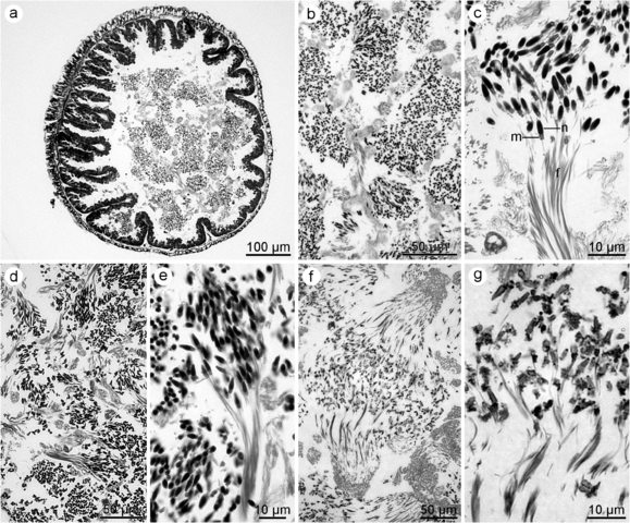
English: FIGURE 3. Histology of spermatozeugmata and sperm cells of Echinopsolus spp. a–c. E. acutus. a. Semithin cross section in centre of seminiferous tubule. b. Sperm bundles fill central space of seminiferous tubule. c. Longitudinal section through spermatozeugma showing sperm heads, mid-pieces and flagella. d–e. E. splendidus. d. Mass of sperm bundles. e. Longitudinal section through spermatozeugma. f–g. E. mollis. f. Mass of sperm bundles. g. Longitudinal section through spermatozeugma with poor structure preservation (alcohol conservation, no aldehyde fixation). f—flagellum; m—mid-piece; n—nucleus. |
| Date | |
| Source | Bohn, J.M., Heß, M. 2014. The Antarctic holothurian genus Echinopsolus Gutt, 1990 (Dendrochirotida, Cucumariidae): brood pouches, spermatozoa, spermatozeugmata and taxonomic implications. Zootaxa 3841(4): 573–591. https://doi.org/10.11646/zootaxa.3841.4.7 |
| Author | Bohn & Heß (2014) |
| Permission (Reusing this file) |
This file is licensed under the Creative Commons Attribution 3.0 Unported license.
|
File history
Click on a date/time to view the file as it appeared at that time.
| Date/Time | Thumbnail | Dimensions | User | Comment | |
|---|---|---|---|---|---|
| current | 17:47, 20 July 2023 | | 1,980 × 1,643 (3.72 MB) | Christian Ferrer (talk | contribs) | {{Information | description = {{en|1=FIGURE 3. Histology of spermatozeugmata and sperm cells of ''Echinopsolus'' spp. a–c. ''E. acutus''. a. Semithin cross section in centre of seminiferous tubule. b. Sperm bundles fill central space of seminiferous tubule. c. Longitudinal section through spermatozeugma showing sperm heads, mid-pieces and flagella. d–e. ''E. splendidus''. d. Mass of sperm bundles. e. Longitudinal section through spermatozeugma. f–g. ''E. mollis''. f. Mass of sperm bundles. g... |
You cannot overwrite this file.
File usage on Commons
The following 2 pages use this file:
_Figure_3.png&oldid=785161768)