File:EB1911 Peripatus - Series of Embryos.jpg

Original file (1,011 × 723 pixels, file size: 218 KB, MIME type: image/jpeg)
Captions
Captions
Summary
[edit]| Description |
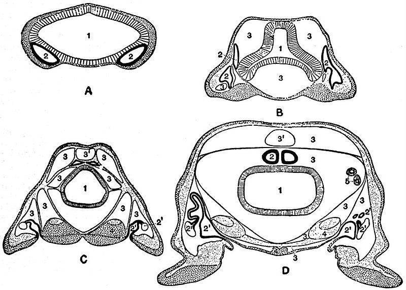
English: A series of diagrams of transverse sections through Peripatus embryos
to show the relations of the coelom at successive stages. See legend below. |
|||
| Date | published 1911 | |||
| Source | “Peripatus,” Encyclopædia Britannica (11th ed.), v. 21, 1911, p. 167, fig. 12. | |||
| Author | After Sedgwick. | |||
| Permission (Reusing this file) |
|

A, Early stage; no trace of the vascular space; endoderm and ectoderm in contact.
B, Endoderm has separated from the dorsal and ventral ectoderm. The somite is represented as having divided on the left side into a dorsal and ventral portion.
C, The haemocoele (3) has become divided up into a number of spaces, the arrangement of which is unimportant. The dorsal part of the somite has travelled dorsalwards, and now constitutes a small space (triangular in section) just dorsal to the gut. The ventral portion (2) has assumed a tubular character, and has acquired an external opening. The internal vesicle is already indicated, and is shown in the diagram by the thinner black line: 1, gut; 2, somite; 2′, nephridial part of coelom; 3, haemocoele; 3′, part of haemocoele which will form the heart—the part of the haemocoele on each side of this will form the pericardium; 4, nerve-cord; 4, slime glands.
D represents the conditions at the time of birth. The coelom is represented as surrounded by a thick black line, except in the part which forms the internal vesicle of the nephridium.
File history
Click on a date/time to view the file as it appeared at that time.
| Date/Time | Thumbnail | Dimensions | User | Comment | |
|---|---|---|---|---|---|
| current | 17:51, 15 May 2019 | | 1,011 × 723 (218 KB) | Bob Burkhardt (talk | contribs) | {{Information |description ={{en|1=A series of diagrams of transverse sections through ''Peripatus'' embryos to show the relations of the coelom at successive stages. See legend below.}} |date =published 1911 |source =“Peripatus,” ''Encyclopædia Britannica'' (11th ed.), v. 21, 1911, p. 167, fig. 12. |author =After Sedgwick. |permission ={{PD-Britannica}} }} {{en|Legend:}} A, Early stage; no trace of the vascular space; endoderm and ectoderm in contact. B, Endode... |
You cannot overwrite this file.
File usage on Commons
There are no pages that use this file.
File usage on other wikis
The following other wikis use this file:
- Usage on en.wikisource.org