File:Dirofilaria repens occular cyst.jpg
Dirofilaria_repens_occular_cyst.jpg (600 × 550 pixels, file size: 56 KB, MIME type: image/jpeg)
Captions
Captions
| Description |
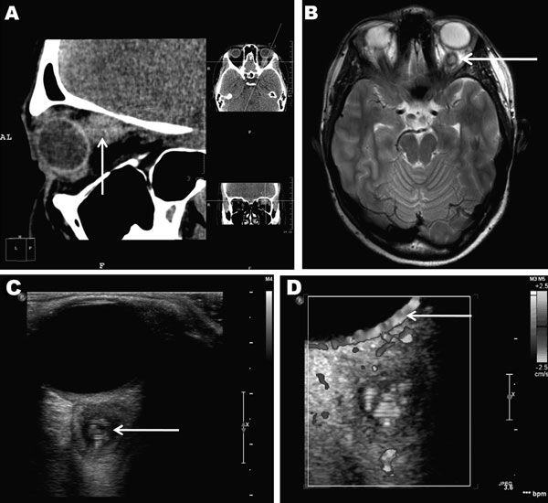
English: Retroocular nodule of a Dirofilaria repens worm detected in a 20-year-old woman, Rostov-na-Donu, Russia. The cyst (arrows) is shown by computed tomography scan (A) and magnetic resonance imaging (B). Ultrasonography image (C) shows a worm-like structure inside the cyst (arrow), and color Doppler imaging (D) shows marginal vascularization of the lesion). |
|||
| Date | Unknown date, published in February 2013 | |||
| Source | Emerging Infectious Diseases, Volume 19, Number 2—February 2013 , http://wwwnc.cdc.gov/eid/article/19/2/12-1388-f1.htm | |||
| Author | Boris Ilyasov, Vladimir Kartashev, Nikolay Bastrikov, Rodrigo Morchón, Javier González-Miguel, and Fernando Simón | |||
| Permission (Reusing this file) |
Copyright and Disclaimers The opinions expressed by authors contributing to this journal do not necessarily reflect the opinions of CDC or the institutions with which the authors are affiliated. Emerging Infectious Diseases is published by the Centers for Disease Control and Prevention, a U.S. Government agency. Therefore, all materials published in Emerging Infectious Diseases are in the public domain and can be used without permission. Proper citation, however, is required. Use of trade names is for identification only and does not imply endorsement by the Public Health Service or by the U.S. Department of Health and Human Services. Emerging Infectious Diseases is printed on acid free paper that meets the requirements of ANSI/NISO 239.48-1992 (Permanence of Paper) |
File history
Click on a date/time to view the file as it appeared at that time.
| Date/Time | Thumbnail | Dimensions | User | Comment | |
|---|---|---|---|---|---|
| current | 03:24, 31 January 2013 | | 600 × 550 (56 KB) | Oaktree b (talk | contribs) | {{Information |Description ={{en|1=Retroocular nodule of a Dirofilaria repens worm detected in a 20-year-old woman, Rostov-na-Donu, Russia. The cyst (arrows) is shown by computed tomography scan (A) and magnetic resonance imaging (B). Ultrasonograph... |
You cannot overwrite this file.
File usage on Commons
There are no pages that use this file.
File usage on other wikis
The following other wikis use this file:
- Usage on en.wikipedia.org