File:Dinophysis tripos.png
Jump to navigation
Jump to search

Size of this preview: 500 × 599 pixels. Other resolutions: 200 × 240 pixels | 400 × 480 pixels | 641 × 768 pixels | 854 × 1,024 pixels | 2,056 × 2,464 pixels.
Original file (2,056 × 2,464 pixels, file size: 3.32 MB, MIME type: image/png)
Captions
Captions
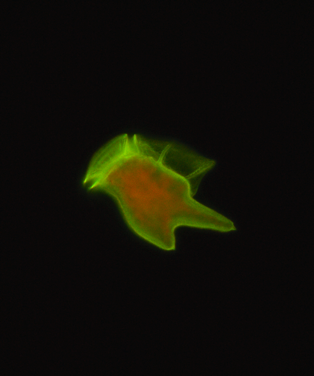
Cell of the dinoflagellate Dinophysis tripos observed with an epifluorescence microscope (magnification x640).
Summary
[edit]
| Description |
Français : Cellule du dinoflagellé Dinophysis tripos observée au microscope optique à épifluorescence (grossissement x640). Cellule issue d'une culture maintenue au Laboratoire PHYSALG de l'IFREMER, Nantes, France. La thèque (paroi) du dinoflagellé, faite de cellulose, a été colorée au Direct Yellow 96, qui fluoresce en vert-jaune. Les chloroplastes dont la chlorophylle a fluoresce en rouge sont visibles à l'intérieur de la cellule. Photographie de Victor Pochic.
English: Cell of the dinoflagellate Dinophysis tripos observed with an epifluorescence microscope (magnification x640). Cell from a culture maintained at the IFREMER PHYSALG lab, Nantes, France. The theca (cell wall) of the dinoflagellate, made of cellulose, was colored with the fluorochrome Direct Yellow 96, that fluoresces in yellow-green. The chloroplasts with chlorophyll a fluorescing in red are visible inside the cell. Photograph by Victor Pochic. English: Cell of the dinoflagellate Dinophysis tripos observed with an epifluorescence microscope (magnification x640). Cell from a culture maintained at the IFREMER PHYSALG lab, Nantes, France. The theca (cell wall) of the dinoflagellate, made of cellulose, was colored with the fluorochrome Direct Yellow 96, that fluoresces in yellow-green. The chloroplasts with chlorophyll a fluorescing in red are visible inside the cell. Photograph by Victor Pochic. |
| Date | |
| Source | Own work |
| Author | Phycoplankton |
Licensing
[edit]
I, the copyright holder of this work, hereby publish it under the following license:
This file is licensed under the Creative Commons Attribution-Share Alike 4.0 International license.
- You are free:
- to share – to copy, distribute and transmit the work
- to remix – to adapt the work
- Under the following conditions:
- attribution – You must give appropriate credit, provide a link to the license, and indicate if changes were made. You may do so in any reasonable manner, but not in any way that suggests the licensor endorses you or your use.
- share alike – If you remix, transform, or build upon the material, you must distribute your contributions under the same or compatible license as the original.
| This file was uploaded as part of Wiki Science Competition 2023. |
File history
Click on a date/time to view the file as it appeared at that time.
| Date/Time | Thumbnail | Dimensions | User | Comment | |
|---|---|---|---|---|---|
| current | 13:25, 22 November 2023 | | 2,056 × 2,464 (3.32 MB) | Phycoplankton (talk | contribs) | Uploaded own work with UploadWizard |
You cannot overwrite this file.
File usage on Commons
There are no pages that use this file.