File:Diagnostics-11-02205-g001.png
Jump to navigation
Jump to search

Size of this preview: 800 × 518 pixels. Other resolutions: 320 × 207 pixels | 640 × 414 pixels | 1,024 × 663 pixels | 1,280 × 829 pixels | 2,560 × 1,657 pixels | 4,270 × 2,764 pixels.
Original file (4,270 × 2,764 pixels, file size: 2.79 MB, MIME type: image/png)
Captions
Captions
Acute Hepatic Porphyrias
Summary
[edit]
| Description |
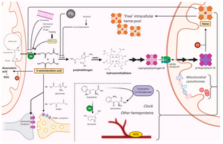
English: Figure 1. Mechanisms of neuronal damage in acute hepatic porphyrias. ALAS condenses glycine and succinyl-CoA into ALA, in the first step of the biosynthesis of heme. After an initial series of reactions in the cytosol, coproporphyrin III is imported in mitochondria by ABCB6, a homodimeric porphyrin transporter located in the outer mitochondrial membrane. ALAS is induced by porphyrinogenic stimuli (e.g., fasting, alcohol, or certain drugs) which supposedly induce an increased metabolic demand for heme. In particular, fasting induces ALAS1 expression via the peroxisome proliferator-activated receptor γ coactivator 1α (PGC-1α). Lead and succinyl acetone cause a porphyria-like picture since they inhibit ALAD. Acute intermittent porphyria, the most common AHP, is an autosomal dominant disease caused by an abnormal function of HMBS. ALA and (in most cases) PBG accumulate in patients with acute porphyrias during neurovisceral attacks. Givosiran, a siRNA-based drug for the treatment of AHPs, acts by impairing ALAS mRNA translation in the liver. Among other toxic effects, ALA undergoes auto-enolization to yield the highly reactive dioxovaleric acid (DOVA) and other oxidant species; it also interferes with GABA and glutamate receptors. Lack of heme has pleiotropic effects on cytochromes, nitric oxide synthases, tryptophan 2,3-dioxygenase, and several other hemeprotein; it may also affect the regulatory functions of the intracellular “free” heme pool. Pyridoxal phosphate figures among the factors involved in this highly connected network of reactions. Other possible mechanisms of neuronal damage are described in the text. ABCB6, ATP-binding cassette transporter B6; ALA, δ-aminolevulinic acid; ALAD, ALA dehydratase; ALAS, ALA synthase; B6, pyridoxal phosphate; Fe, iron; GABA, γ-aminobutyric acid; HMBS, hydroxymethylbilane synthase; NOS, nitric oxide synthase; Pb, lead. Created with BioRender.com (last accessed date: 22 November 2021). |
| Date | |
| Source | https://www.mdpi.com/2075-4418/11/12/2205 |
| Author | Ricci, A.; Di Pierro, E.; Marcacci, M.; Ventura, P. |
Licensing
[edit]
This file is licensed under the Creative Commons Attribution 4.0 International license.
- You are free:
- to share – to copy, distribute and transmit the work
- to remix – to adapt the work
- Under the following conditions:
- attribution – You must give appropriate credit, provide a link to the license, and indicate if changes were made. You may do so in any reasonable manner, but not in any way that suggests the licensor endorses you or your use.
File history
Click on a date/time to view the file as it appeared at that time.
| Date/Time | Thumbnail | Dimensions | User | Comment | |
|---|---|---|---|---|---|
| current | 21:22, 13 June 2024 | | 4,270 × 2,764 (2.79 MB) | Ozzie10aaaa (talk | contribs) | Uploaded a work by Ricci, A.; Di Pierro, E.; Marcacci, M.; Ventura, P. from https://www.mdpi.com/2075-4418/11/12/2205 with UploadWizard |
You cannot overwrite this file.
File usage on Commons
There are no pages that use this file.