File:Delamination of the cephalic and trunk neural crest cells.jpg

Original file (2,963 × 2,818 pixels, file size: 873 KB, MIME type: image/jpeg)
Captions
Captions
Summary
[edit]
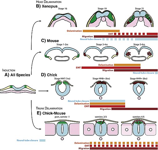
| Description | Fig. 1. Delamination of the cephalic and trunk neural crest cells. (A) Basic organization of the dorsal region of a vertebrate embryo at early neurula stage. NC cells (green) are induced at the border of the open neural plate (blue). (B) Xenopus cephalic NC cells separate from the open neural plate and the sensory layer of the ectoderm between stages 16 and 18 and start migrating as a cohesive group (stage 19). While migration proceeds, NC cells become progressively more mesenchymal (red cells). Based on Slug and Foxd3 expressions on histological sections and electron microscopy after Schroeder (1970). (C) Delamination of mouse cephalic NC cells starts at open neural plate stage. NC cells undergo an EMT, delaminate and start migrating within a few hours. Modified after Nichols (1987). (D) Delamination of chick cephalic NC cells involves a massive EMT. All cells delaminate at once and start migrating soon after. Based on the dynamic of Ets1 expression in the mesencephalon. Modified after Theveneau et al. (2007) (E) Delamination of chick/mouse rostral trunk NC cells. Premigratory NC cells that are located in the dorsal part of the closed neural tube face the presomitic mesoderm (psm). Delamination starts in front of the second/third newly formed somites. NC cells undergo EMT one by one, delaminate in a dripping fashion and start migrating as soon as they leave the neural tube. Modified after Kos et al. (2001) and Sela-Donenfeld and Kalcheim (1999). Note that neural tube closure and NC delamination are not synchronized across species. Also note that the timing of delamination and EMT do not necessarily coincide. Premigratory NC territory and NC cells are shown in green, red round cells represent mesenchymal NC cells or NC cells undergoing EMT. The neural plate/tube is in blue, non-neural ectoderm and the sensory layer of the ectoderm are in yellow, mesoderm and its derivatives are in pink. |
| Date | |
| Source |
https://www.sciencedirect.com/science/article/pii/S0012160611014692?via%3Dihub Neural crest delamination and migration: From epithelium-to-mesenchyme transition to collective cell migration, Developmental Biology, Volume 366, Issue 1, 2012, Pages 34-54, ISSN 0012-1606, https://doi.org/10.1016/j.ydbio.2011.12.041. (https://www.sciencedirect.com/science/article/pii/S0012160611014692) |
| Author | Eric Theveneau, Roberto Mayor, |

|
This file, which was originally posted to an external website, has not yet been reviewed by an administrator or reviewer to confirm that the above license is valid. See Category:License review needed for further instructions.
|
https://s100.copyright.com/AppDispatchServlet?publisherName=ELS&contentID=S0012160611014692&orderBeanReset=true This is an open access article distributed under the terms of the Creative Commons CC-BY license, which permits unrestricted use, distribution, and reproduction in any medium, provided the original work is properly cited. You are not required to obtain permission to reuse this article.
Licensing
[edit]
- You are free:
- to share – to copy, distribute and transmit the work
- to remix – to adapt the work
- Under the following conditions:
- attribution – You must give appropriate credit, provide a link to the license, and indicate if changes were made. You may do so in any reasonable manner, but not in any way that suggests the licensor endorses you or your use.
File history
Click on a date/time to view the file as it appeared at that time.
| Date/Time | Thumbnail | Dimensions | User | Comment | |
|---|---|---|---|---|---|
| current | 19:15, 2 June 2024 | | 2,963 × 2,818 (873 KB) | Rasbak (talk | contribs) | {{Information |description=Fig. 1. Delamination of the cephalic and trunk neural crest cells. (A) Basic organization of the dorsal region of a vertebrate embryo at early neurula stage. NC cells (green) are induced at the border of the open neural plate (blue). (B) Xenopus cephalic NC cells separate from the open neural plate and the sensory layer of the ectoderm between stages 16 and 18 and start migrating as a cohesive group (stage 19). While migration proceeds, NC cells become progressively... |
You cannot overwrite this file.
File usage on Commons
There are no pages that use this file.
File usage on other wikis
The following other wikis use this file:
- Usage on nl.wikipedia.org