File:Corbicula fluminea veligers and early pediveligers.jpg
Jump to navigation
Jump to search
Size of this preview: 486 × 600 pixels. Other resolutions: 194 × 240 pixels | 389 × 480 pixels | 622 × 768 pixels | 1,025 × 1,265 pixels.
Original file (1,025 × 1,265 pixels, file size: 1.27 MB, MIME type: image/jpeg)
Captions
Captions
Add a one-line explanation of what this file represents
Summary
[edit]
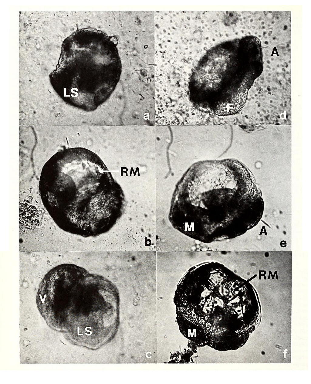
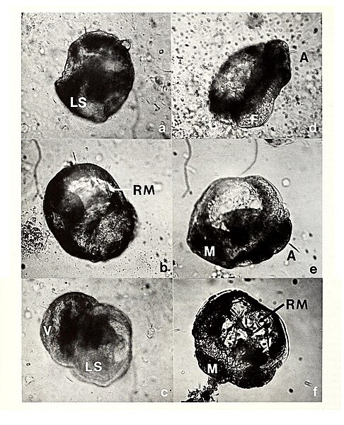
| Description | Fig. 8 a-f. Photomicrographs of veligers and early pediveligers of Corbicula fluminea. (a), (b), (c) veligers. (b) especially, shows swollen aspect of a veliger in osmotic distress after being exposed to river water. (d), (e), (f) pediveligers. (d) velum and foot extended, mantle retracted. (e) velum extended; mantle extended in posterior region. (f) velum extended; obscuring extended foot. A, apical cilia; F, foot, LS, larval shell valves; RM, velar retractor muscle; V, velum. Horizontal field width = 255 um. |
| Date | |
| Source | https://archive.org/details/americanmal4519861987amer/page/n77/mode/1up?view=theater |
| Author |
Licensing
[edit]
This work is in the public domain because it was published in the United States between 1978 and March 1, 1989 without a copyright notice, and its copyright was not subsequently registered with the U.S. Copyright Office within 5 years. Unless its author has been dead for several years, it is copyrighted in the countries or areas that do not apply the rule of the shorter term for US works, such as Canada (50 pma), Mainland China (50 pma, not Hong Kong or Macau), Germany (70 pma), Mexico (100 pma), Switzerland (70 pma), and other countries with individual treaties. See this page for further explanation.
|
 |

|
This file, which was originally posted to an external website, has not yet been reviewed by an administrator or reviewer to confirm that the above license is valid. See Category:License review needed for further instructions.
|
[[Category:Plankton]
File history
Click on a date/time to view the file as it appeared at that time.
| Date/Time | Thumbnail | Dimensions | User | Comment | |
|---|---|---|---|---|---|
| current | 21:51, 22 March 2024 | | 1,025 × 1,265 (1.27 MB) | Rasbak (talk | contribs) | {{information |description=Fig. 8 a-f. Photomicrographs of veligers and early pediveligers of Corbicula fluminea. (a), (b), (c) veligers. (b) especially, shows swollen aspect of a veliger in osmotic distress after being exposed to river water. (d), (e), (f) pediveligers. (d) velum and foot extended, mantle retracted. (e) velum extended; mantle extended in posterior region. (f) velum extended; obscuring extended foot. A, apical cilia; F, foot, LS, larval shell valves; RM, velar retractor muscl... |
You cannot overwrite this file.
File usage on Commons
There are no pages that use this file.