File:Congenital occipital encephalocele in an infant Wellcome L0062875.jpg
Jump to navigation
Jump to search
Size of this preview: 800 × 505 pixels. Other resolutions: 320 × 202 pixels | 640 × 404 pixels | 1,024 × 647 pixels | 1,280 × 809 pixels | 2,560 × 1,617 pixels | 5,555 × 3,509 pixels.
Original file (5,555 × 3,509 pixels, file size: 1.99 MB, MIME type: image/jpeg)
Captions
Captions
Medical photograph taken to illustrate the pathology of a medical condition
Summary
[edit]
| Congenital occipital encephalocele in an infant
( |
|||||||||||||||||||||||
|---|---|---|---|---|---|---|---|---|---|---|---|---|---|---|---|---|---|---|---|---|---|---|---|
| Title |
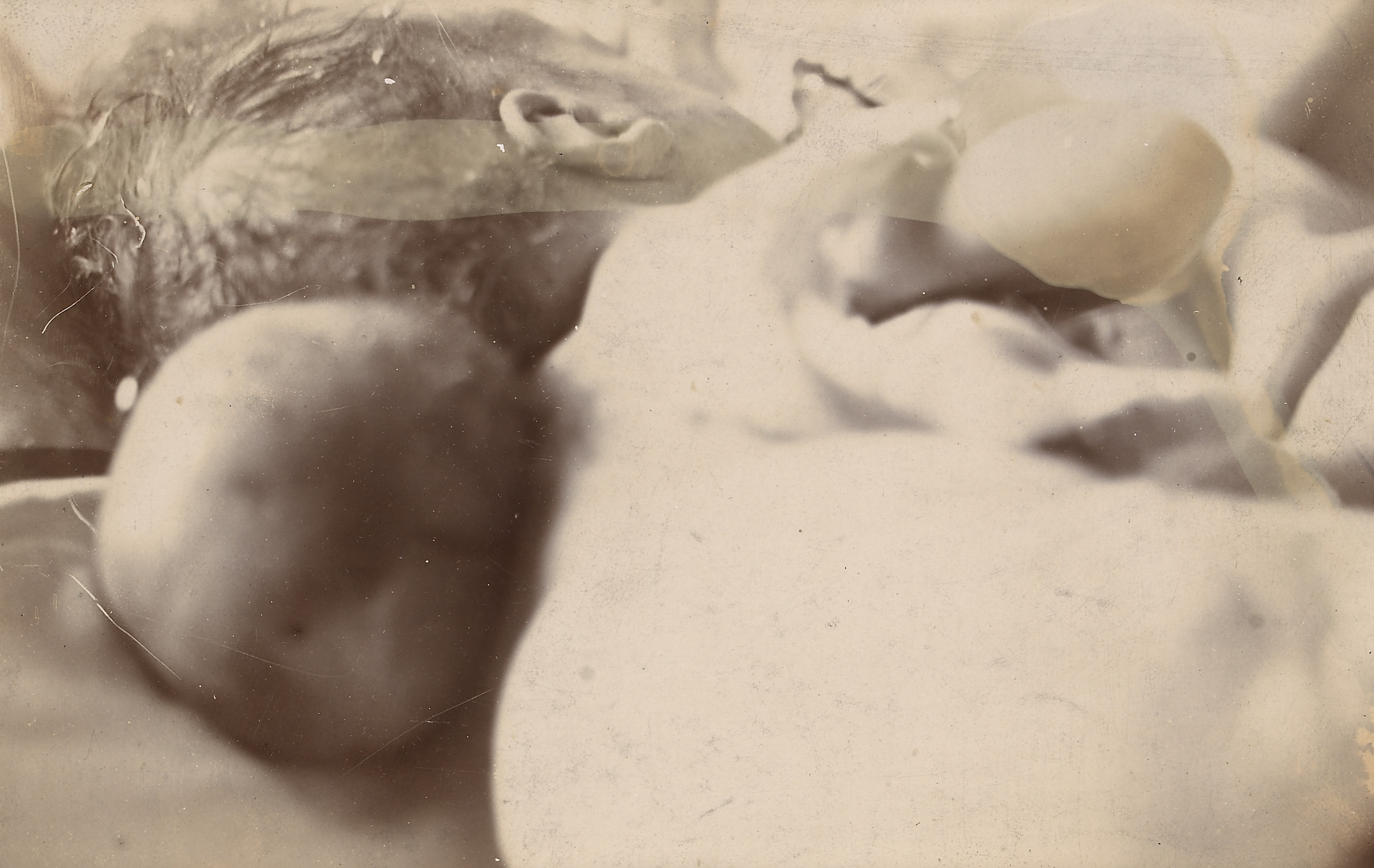
Congenital occipital encephalocele in an infant |
||||||||||||||||||||||
| Description |
Black and white photograph showing a congenital occipital encephalocele. The patient was an infant of two months. The encephalocele was excised, but the child died shortly after the operation. The autopsy showed that the herniated brain-matter came through a hole of about half an inch in diameter, situated two inches below the upper extremity of the occipital bone. The cavity of the encephalocele communicated with the lateral ventricles. The foramen magnum was partially deficient behind, as were the arches of the atlas and axis vetrebrae. The cerebellum lay in a funnel shaped depression formed by this deficiency. |
||||||||||||||||||||||
| Date | 19 Aug 1905 | ||||||||||||||||||||||
| Collection |
|
||||||||||||||||||||||
| Credit line | This is an image of an item held by Barts Health NHS Trust Archives. | ||||||||||||||||||||||
| References |
Barts Health NHS Trust Archives SBHB/MU/14/30/41
|
||||||||||||||||||||||
| Source/Photographer | https://www.calmview.co.uk/BartsHealth/CalmView/Record.aspx?src=CalmView.Catalog&id=SBHB%2fMU%2f14%2f30%2f41&pos=1 | ||||||||||||||||||||||
Licensing
[edit]
This file is licensed under the Creative Commons Attribution 4.0 International license.
- You are free:
- to share – to copy, distribute and transmit the work
- to remix – to adapt the work
- Under the following conditions:
- attribution – You must give appropriate credit, provide a link to the license, and indicate if changes were made. You may do so in any reasonable manner, but not in any way that suggests the licensor endorses you or your use.
File history
Click on a date/time to view the file as it appeared at that time.
| Date/Time | Thumbnail | Dimensions | User | Comment | |
|---|---|---|---|---|---|
| current | 07:53, 18 October 2014 | | 5,555 × 3,509 (1.99 MB) | Fæ (talk | contribs) | =={{int:filedesc}}== {{Artwork |artist = |author = |title = Congenital occipital encephalocele in an infant |description = Black and white photograph showing a congenital occipital encephalocele. The patient... |
You cannot overwrite this file.
File usage on Commons
The following page uses this file:
File usage on other wikis
The following other wikis use this file:
- Usage on es.wikipedia.org