File:Brucella melitensis flagellum (fig. 2).png
Jump to navigation
Jump to search
Size of this preview: 688 × 600 pixels. Other resolutions: 275 × 240 pixels | 551 × 480 pixels | 881 × 768 pixels | 1,200 × 1,046 pixels.
Original file (1,200 × 1,046 pixels, file size: 760 KB, MIME type: image/png)
Captions
Captions
Add a one-line explanation of what this file represents
Summary
[edit].png&action=edit§ion=1)
| Description |
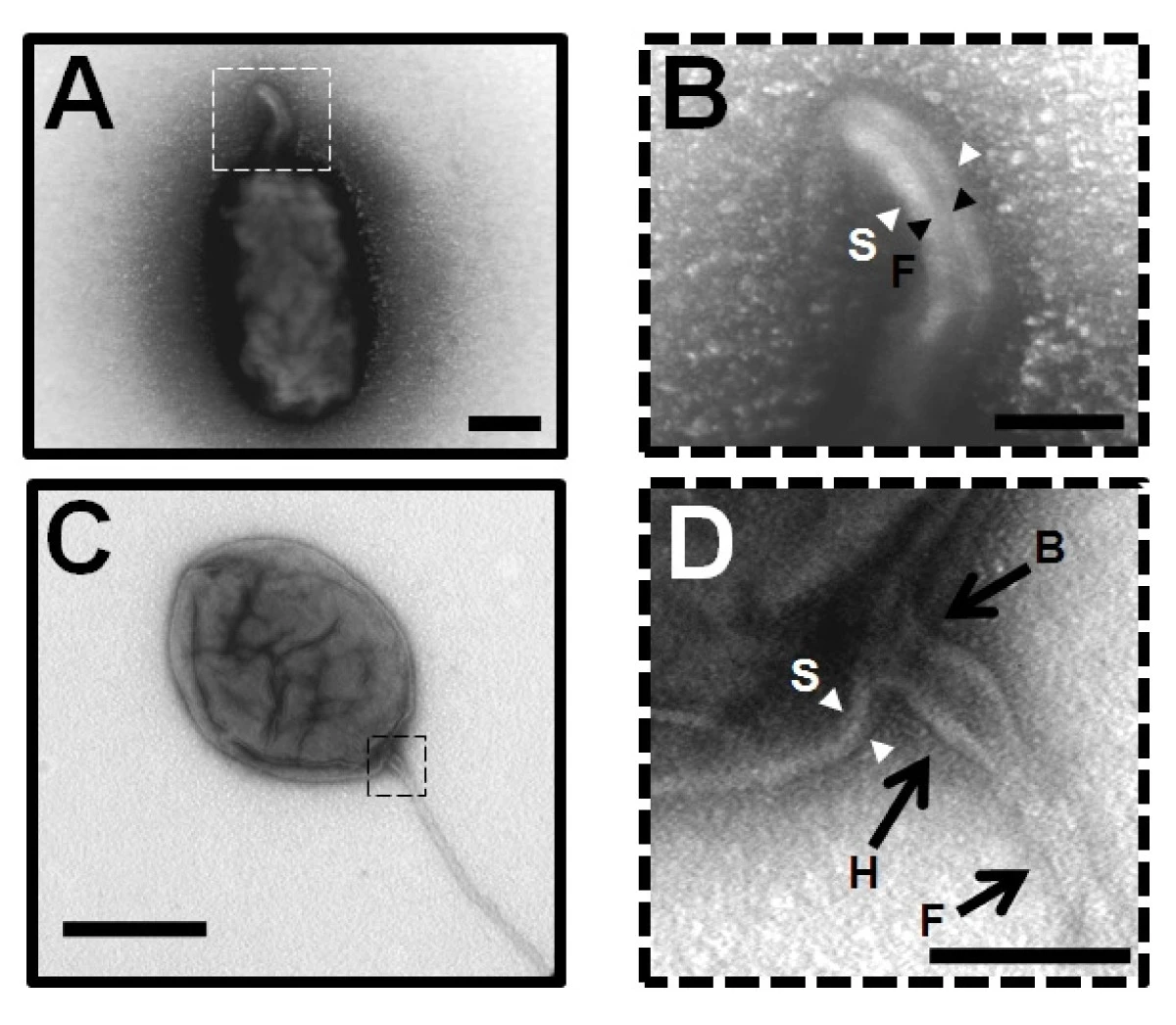
English: A and B – negative-staining EM images of the sheathed polar flagellum of Brucella melitensis stained with uranyl acetate 2%. B – the central filament is indicated by black arrowheads with F and the sheath by white arrowheads with S. Bar, 200 nm. C and D – capture of the basal region of the flagellum. The image shows the basal body region (B with black arrow), the hook (H with black arrow) and is finished by the filament (F with black arrow). The structure is surrounded by a sheath extended from the outer membrane (S, white arrowheads). Bar, 100 nm. The images B and D are enlarged from the dotted square in image A and C respectively. |
| Date | |
| Source | Ferooz J., Letesson J. J. (2010). "Morphological analysis of the sheathed flagellum of Brucella melitensis". BMC Res Notes 3: 333. DOI:10.1186/1756-0500-3-333. |
| Author | Jonathan Ferooz, Jean-Jacques Letesson |
Licensing
[edit].png&action=edit§ion=2)
This file is licensed under the Creative Commons Attribution-Share Alike 2.0 Generic license.
- You are free:
- to share – to copy, distribute and transmit the work
- to remix – to adapt the work
- Under the following conditions:
- attribution – You must give appropriate credit, provide a link to the license, and indicate if changes were made. You may do so in any reasonable manner, but not in any way that suggests the licensor endorses you or your use.
- share alike – If you remix, transform, or build upon the material, you must distribute your contributions under the same or compatible license as the original.
File history
Click on a date/time to view the file as it appeared at that time.
| Date/Time | Thumbnail | Dimensions | User | Comment | |
|---|---|---|---|---|---|
| current | 22:29, 21 January 2021 | | 1,200 × 1,046 (760 KB) | Thinknot (talk | contribs) | {{Information |Description= {{en|1='''A''' and '''B''' – negative-staining EM images of the sheathed polar flagellum of ''Brucella melitensis'' stained with uranyl acetate 2%. '''B''' – the central filament is indicated by black arrowheads with ''F'' and the sheath by white arrowheads with ''S''. Bar, 200 nm. '''C''' and '''D''' – capture of the basal region of the flagellum. The image shows the basal body region (''B'' with black arrow), the hook (''H'' with black arrow) and is finished by t... |
You cannot overwrite this file.
File usage on Commons
There are no pages that use this file.
File usage on other wikis
The following other wikis use this file:
- Usage on ru.wikipedia.org
- Usage on tr.wikipedia.org
.png&oldid=835491650)