File:Brucella melitensis flagellum (fig. 1).png
Jump to navigation
Jump to search

Size of this preview: 800 × 321 pixels. Other resolutions: 320 × 128 pixels | 640 × 257 pixels | 1,200 × 481 pixels.
Original file (1,200 × 481 pixels, file size: 362 KB, MIME type: image/png)
Captions
Captions
Add a one-line explanation of what this file represents
Summary
[edit].png&action=edit§ion=1)
| Description |
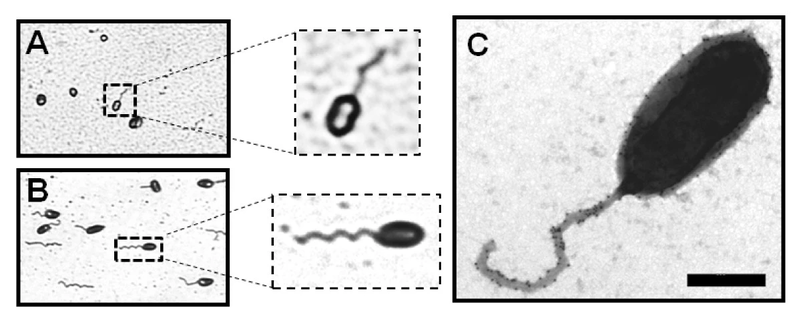
English: Ryu staining of Brucella melitensis (A) and Caulobacter crescentus (B) observed by phase-contrast microscopy. The samples were treated by the Ryu staining method. A flagellated bacterium is enlarged in a dotted square. (C) Negative-staining EM images of the sheathed polar flagellum of B. melitensis stained with uranyl acetate 2% and labeled with anti-LPS antibody conjugated to ± 15 nm gold particles. Bar, 500 nm. |
| Date | |
| Source | Ferooz J., Letesson J. J. (2010). "Morphological analysis of the sheathed flagellum of Brucella melitensis". BMC Res Notes 3: 333. DOI:10.1186/1756-0500-3-333. |
| Author | Jonathan Ferooz, Jean-Jacques Letesson |
Licensing
[edit].png&action=edit§ion=2)
This file is licensed under the Creative Commons Attribution-Share Alike 2.0 Generic license.
- You are free:
- to share – to copy, distribute and transmit the work
- to remix – to adapt the work
- Under the following conditions:
- attribution – You must give appropriate credit, provide a link to the license, and indicate if changes were made. You may do so in any reasonable manner, but not in any way that suggests the licensor endorses you or your use.
- share alike – If you remix, transform, or build upon the material, you must distribute your contributions under the same or compatible license as the original.
File history
Click on a date/time to view the file as it appeared at that time.
| Date/Time | Thumbnail | Dimensions | User | Comment | |
|---|---|---|---|---|---|
| current | 22:24, 21 January 2021 | 1,200 × 481 (362 KB) | Thinknot (talk | contribs) | {{Information |Description= {{en|1=Ryu staining of ''Brucella melitensis'' ('''A''') and ''Caulobacter crescentus'' ('''B''') observed by phase-contrast microscopy. The samples were treated by the Ryu staining method. A flagellated bacterium is enlarged in a dotted square. ('''C''') Negative-staining EM images of the sheathed polar flagellum of ''B. melitensis'' stained with uranyl acetate 2% and labeled with anti-LPS antibody conjugated to ± 15 nm gold particles. Bar, 500 nm.}} |Source= {{ci... |
You cannot overwrite this file.
File usage on Commons
There are no pages that use this file.
.png&oldid=835491646)