File:Beiträge zur Anatomie der weiblichen Urogenitalorgane des Orang-Utan BHL45530048.jpg
Original file (3,903 × 3,091 pixels, file size: 1,008 KB, MIME type: image/jpeg)
Captions
Captions
Summary
[edit]| Description |
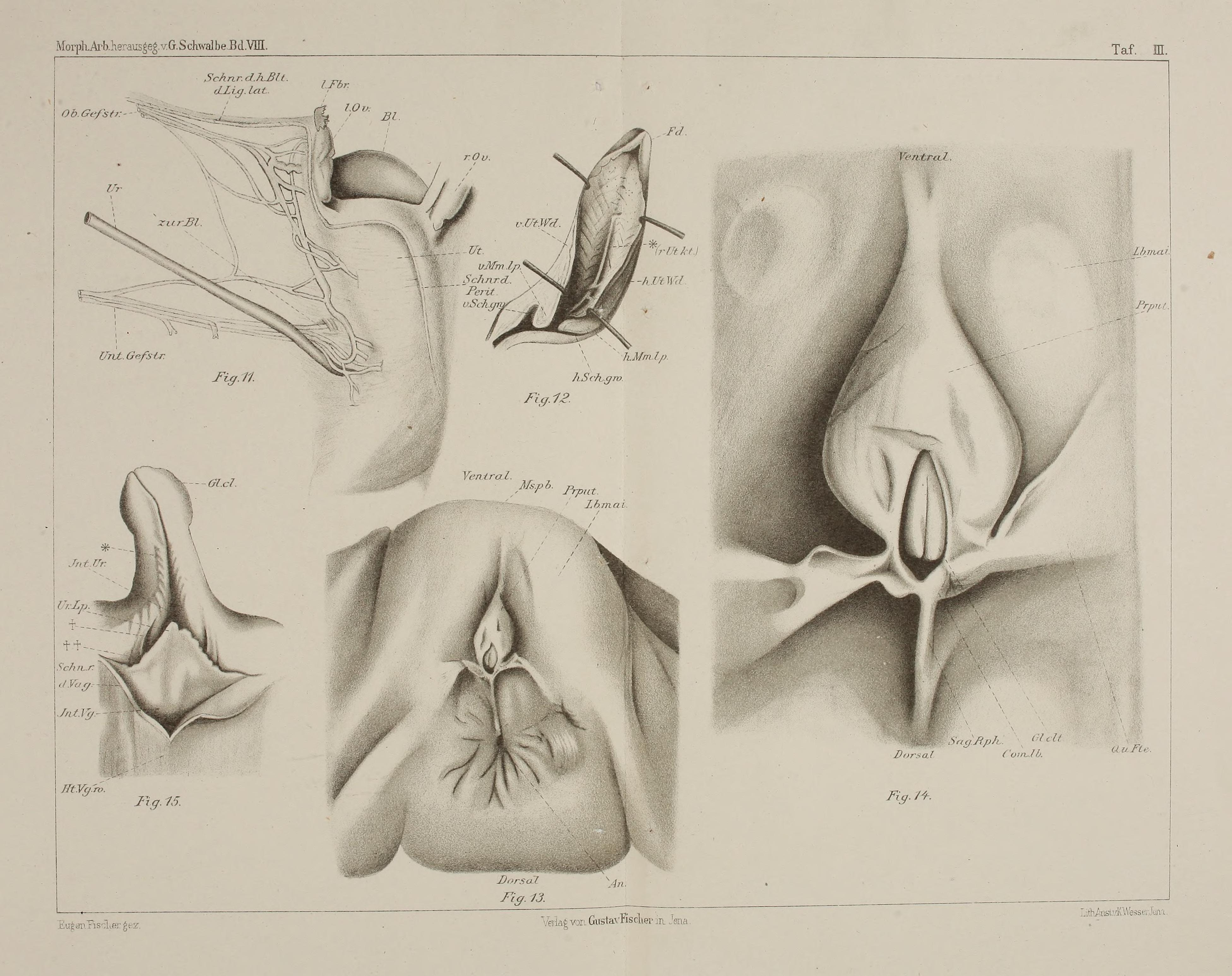
Beiträge zur Anatomie der weiblichen Urogenitalorgane des Orang-Utan. Figur 11. Ansicht des Uterus von hinten nach Entfernung des Bauchfells und Präparation der Gefässe. Etwas vergrössert,
Schnr.d. Perit, = Schnittrand des hinteren Peritonealüber-
zuges des Uterus. Sehnr. dh. Bled. Lg. lat. = Schnitt-
rand des hinteren Blattes des Lig. latum. (+. = Ureter.
Ob. Gef.str. = im lig. lat. verlaufender oberer Gefässstrang;
Unt. Gef. sir. = entsprechender unterer Strang. |
||
| Date | |||
| Source | https://www.biodiversitylibrary.org/pageimage/45530048 | ||
| Author | Fischer, Eugen | ||
| Collection | Ernst Mayr Library of the MCZ, Harvard University | ||
| Page ID | 45530048 | ||
| Item ID | 163298 (Find related Wikimedia Commons images) | ||
| Title ID | 86376 (Find related Wikimedia Commons images) | ||
| BHL Page URL | https://www.biodiversitylibrary.org/page/45530048 | ||
| DOI | 10.5962/bhl.title.86376 | ||
| Page type | Foldout | ||
| Credit | Public domain. The BHL considers that this work is no longer under copyright protection.
|

Licensing
[edit]
This image is in the public domain because it is a mere mechanical scan or photocopy of a public domain original, or – from the available evidence – is so similar to such a scan or photocopy that no copyright protection can be expected to arise. The original itself is in the public domain for the following reason:
This tag is designed for use where there may be a need to assert that any enhancements (eg brightness, contrast, colour-matching, sharpening) are in themselves insufficiently creative to generate a new copyright. It can be used where it is unknown whether any enhancements have been made, as well as when the enhancements are clear but insufficient. For known raw unenhanced scans you can use an appropriate {{PD-old}} tag instead. For usage, see Commons:When to use the PD-scan tag.  | ||||
File history
Click on a date/time to view the file as it appeared at that time.
| Date/Time | Thumbnail | Dimensions | User | Comment | |
|---|---|---|---|---|---|
| current | 22:48, 29 October 2015 | | 3,903 × 3,091 (1,008 KB) | Fæ (talk | contribs) | == {{int:filedesc}} == {{BHL | collection = Ernst Mayr Library of the MCZ, Harvard University | source = http://www.biodiversitylibrary.org/pageimage/45530048 | description = Beiträge zur Anatomie der weiblichen Urogenitalorgane des Orang-Utan. | page... |
You cannot overwrite this file.
File usage on Commons
There are no pages that use this file.