File:Basic mechanical principles of present-day fluoroscopes and Basic mechanical principles of radiographic apparatus.jpg
Original file (1,606 × 2,381 pixels, file size: 1.01 MB, MIME type: image/jpeg)
Captions
Captions
Summary
[edit]| Description |
English: Basic mechanical principles of present-day fluoroscopes and Basic mechanical principles of radiographic apparatus
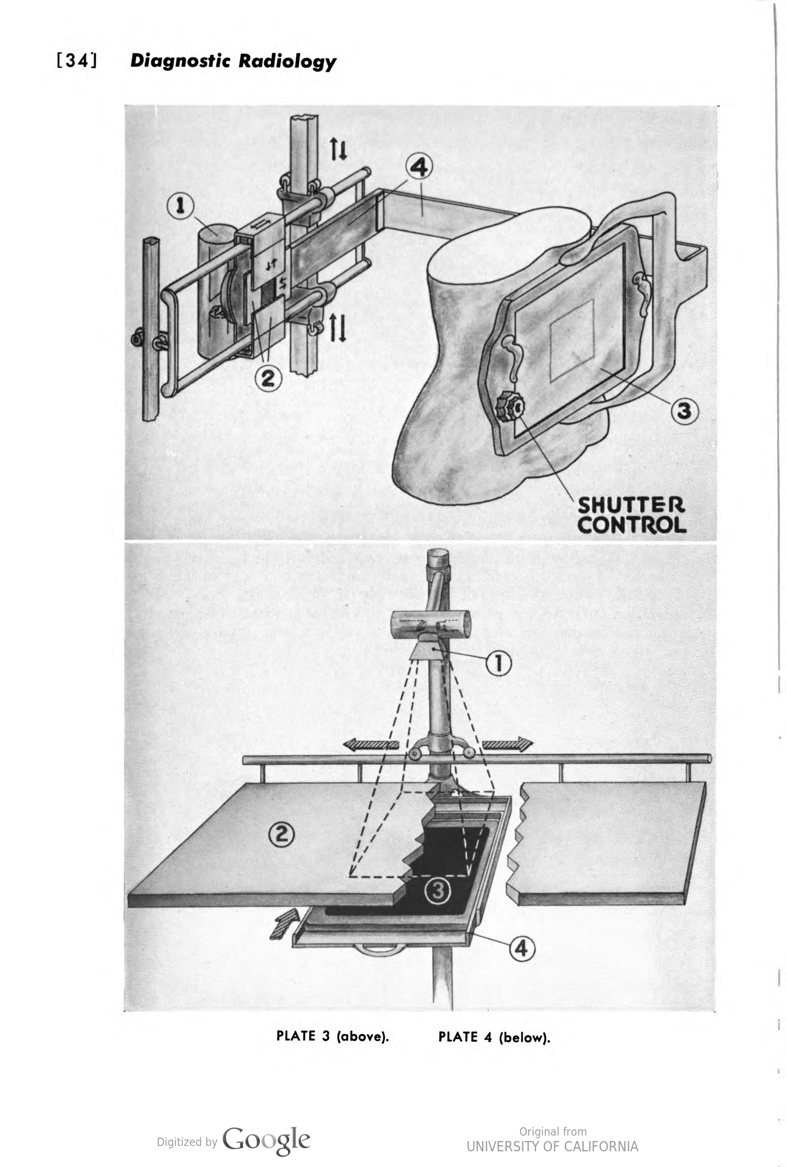
<-PLATE 3.—Basic mechanical principles of present-day fluoroscopes. 1, x-ray tube heavily shielded to contain unwanted radiation, at the same time permitting useful beam to emerge by way of a small window fitted with filters of aluminum to reduce the effect of radiation on patient's skin; 2, heavy sheet-lead shutters to modify size of x-ray beam; 3, transparent lead impregnated glass to protect observer against excessive exposure; 4, rigid arm to which both tube and screen are attached. Counterbalanced and fitted with ball-bearing rollers, entire assembly is freely movable, side to side and up and down, on supporting framework. Fluoroscopes are built in vertical, horizontal and tilting types. PLATE 4.—Basic mechanical principles of radiographic apparatus. Upright mast supported near floor and at table height by sturdy tubular rails, along which it may be shifted on ball-bearing rollers; an arm, adjustable along mast above table, carrying x-ray tube in protective shield; x-ray-proof diaphragm and “come” (1) to limit size of beam to size of film to be ex posed; radiolucent table top on which patient is placed (2); “cassette” or light-proof holder for photographic film (3) lined with fluorescent screens and having “window” opaque to light but transparent to x-rays, and drawer (4) for cassette, which, when closed, places film exactly in path of beam. |
| Date | |
| Source |
Radiology for medical students (1958) Internet Archive identifier: hodges_radiologyformedicalstudents_1958 |
| Author | Hodges, Fred Jenner, 1895- |
| Other versions |


Licensing
[edit]
This work is in the public domain because it was published in the United States between 1929 and 1963, and although there may or may not have been a copyright notice, the copyright was not renewed. For further explanation, see Commons:Hirtle chart and the copyright renewal logs.
|
 |
File history
Click on a date/time to view the file as it appeared at that time.
| Date/Time | Thumbnail | Dimensions | User | Comment | |
|---|---|---|---|---|---|
| current | 13:31, 17 February 2021 | | 1,606 × 2,381 (1.01 MB) | Balkanique (talk | contribs) | Uploaded a work by Hodges, Fred Jenner, 1895- from Radiology for medical students (1958) {{Internet Archive link|1=graphikdergegenw00kapl}} with UploadWizard |
You cannot overwrite this file.
File usage on Commons
The following 2 pages use this file: