File:Bacterial growth on blood agar.jpg
Jump to navigation
Jump to search

Size of this preview: 800 × 450 pixels. Other resolutions: 320 × 180 pixels | 640 × 360 pixels | 1,024 × 576 pixels | 1,280 × 720 pixels | 2,560 × 1,440 pixels | 4,160 × 2,340 pixels.
Original file (4,160 × 2,340 pixels, file size: 2.54 MB, MIME type: image/jpeg)
Captions
Captions
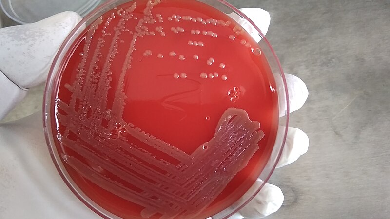
Blood agar is a common type of agar medium used in microbiology to culture and differentiate various bacteria, especially those that are pathogenic. It is an enriched medium that contains nutrients to support the growth of a wide range of bacteria.
Summary
[edit]
| Description |
English: Blood agar is a common type of agar medium used in microbiology to culture and differentiate various bacteria, especially those that are pathogenic. It is an enriched medium that contains nutrients to support the growth of a wide range of bacteria. Bacterial growth on blood agar can reveal several characteristics:Nutrient Richness: Blood agar contains a variety of nutrients, including peptones, beef extract, and agar, making it suitable for the growth of many different bacterial species.Hemolysis: One of the key features of blood agar is its ability to differentiate bacteria based on their hemolytic activity, which is the breakdown of red blood cells in the agar. There are three common types of hemolysis patterns observed on blood agar: Alpha Hemolysis: This is partial hemolysis where bacteria reduce hemoglobin in red blood cells to methemoglobin, causing a greenish discoloration of the agar around the bacterial colonies. Beta Hemolysis: Beta hemolysis is complete hemolysis, and it results in a clear, transparent zone around the bacterial colonies due to the complete lysis of red blood cells. Gamma Hemolysis: In this case, there is no hemolysis, and the agar remains unchanged around the colonies. Colony Morphology: Bacterial colonies on blood agar can have various morphologies, including size, shape, color, and texture. These characteristics can provide initial clues about the type of bacteria present. Streptococci Differentiation: Blood agar is particularly useful for differentiating streptococci (a group of Gram-positive cocci) based on their hemolytic patterns. For example, Streptococcus pyogenes (Group A Streptococcus) exhibits beta hemolysis, while Streptococcus pneumoniae exhibits alpha hemolysis. Identification: The growth of bacteria on blood agar is often followed by additional biochemical tests or techniques such as Gram staining, catalase testing, coagulase testing, and molecular methods to identify and classify the specific bacterial species. |
| Date | |
| Source | Own work |
| Author | Ajay Kumar Chaurasiya |
Licensing
[edit]
I, the copyright holder of this work, hereby publish it under the following license:
This file is licensed under the Creative Commons Attribution-Share Alike 4.0 International license.
- You are free:
- to share – to copy, distribute and transmit the work
- to remix – to adapt the work
- Under the following conditions:
- attribution – You must give appropriate credit, provide a link to the license, and indicate if changes were made. You may do so in any reasonable manner, but not in any way that suggests the licensor endorses you or your use.
- share alike – If you remix, transform, or build upon the material, you must distribute your contributions under the same or compatible license as the original.
File history
Click on a date/time to view the file as it appeared at that time.
| Date/Time | Thumbnail | Dimensions | User | Comment | |
|---|---|---|---|---|---|
| current | 08:48, 2 September 2023 | | 4,160 × 2,340 (2.54 MB) | Ajay Kumar Chaurasiya (talk | contribs) | Uploaded own work with UploadWizard |
You cannot overwrite this file.
File usage on Commons
The following page uses this file: