File:BAX protein 2K7W.png
Jump to navigation
Jump to search
Size of this preview: 618 × 599 pixels. Other resolutions: 248 × 240 pixels | 495 × 480 pixels | 792 × 768 pixels | 1,056 × 1,024 pixels | 1,575 × 1,527 pixels.
Original file (1,575 × 1,527 pixels, file size: 735 KB, MIME type: image/png)
Captions
Captions
Add a one-line explanation of what this file represents
| Description |
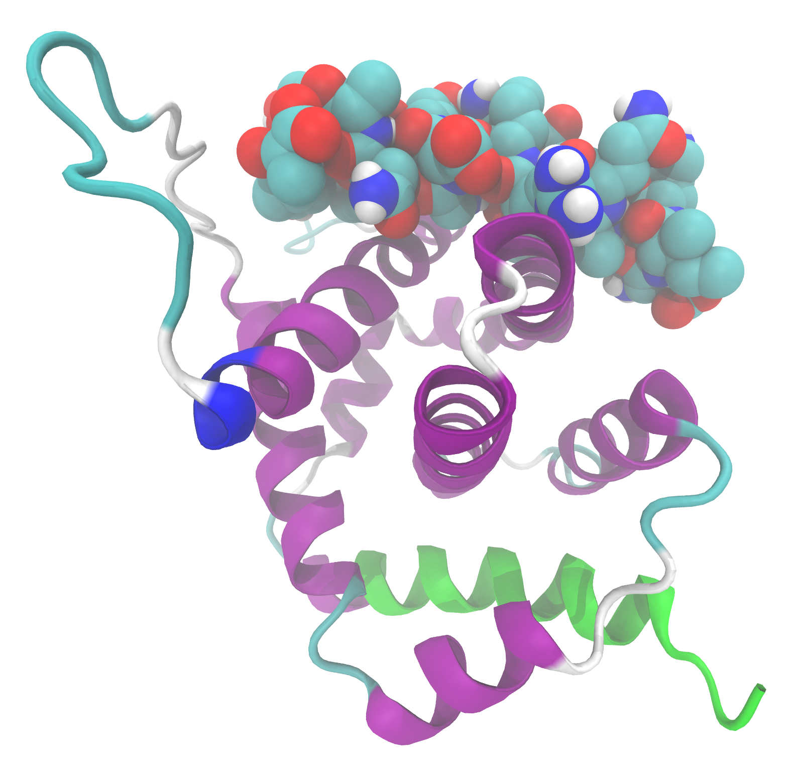
English: Ribbon model of Bax complexed to Bcl2-L-11 BH3-domain (balls). The green residues belong to the transmembrane domain. Ref.: Gavathiotis E, Suzuki M, Davis ML, et al. (October 2008). "BAX activation is initiated at a novel interaction site". Nature 455 (7216): 1076–81. DOI:10.1038/nature07396. PMID 18948948. PMC: 2597110.
This image was created with VMD. |
||
| Date | |||
| Source | Own work | ||
| Author | Ayacop | ||
| Permission (Reusing this file) |
|
File history
Click on a date/time to view the file as it appeared at that time.
| Date/Time | Thumbnail | Dimensions | User | Comment | |
|---|---|---|---|---|---|
| current | 11:47, 4 December 2009 | | 1,575 × 1,527 (735 KB) | Ayacop (talk | contribs) | {{Information |Description={{en|1=Ribbon model of Bax complexed to Bcl2-L-11 BH3-domain (balls). The green residues belong to the transmembrane domain. Ref.: {{cite journal |author=Gavathiotis E, Suzuki M, Davis ML, ''et al.'' |title=BAX activation is ini |
You cannot overwrite this file.
File usage on Commons
There are no pages that use this file.