File:Asteroid body very high mag.jpg
Jump to navigation
Jump to search
Size of this preview: 800 × 533 pixels. Other resolutions: 320 × 213 pixels | 640 × 427 pixels | 1,024 × 683 pixels | 1,280 × 853 pixels | 2,560 × 1,707 pixels | 4,272 × 2,848 pixels.
Original file (4,272 × 2,848 pixels, file size: 2.65 MB, MIME type: image/jpeg)
Captions
Captions
Add a one-line explanation of what this file represents
Summary
[edit]| Description |
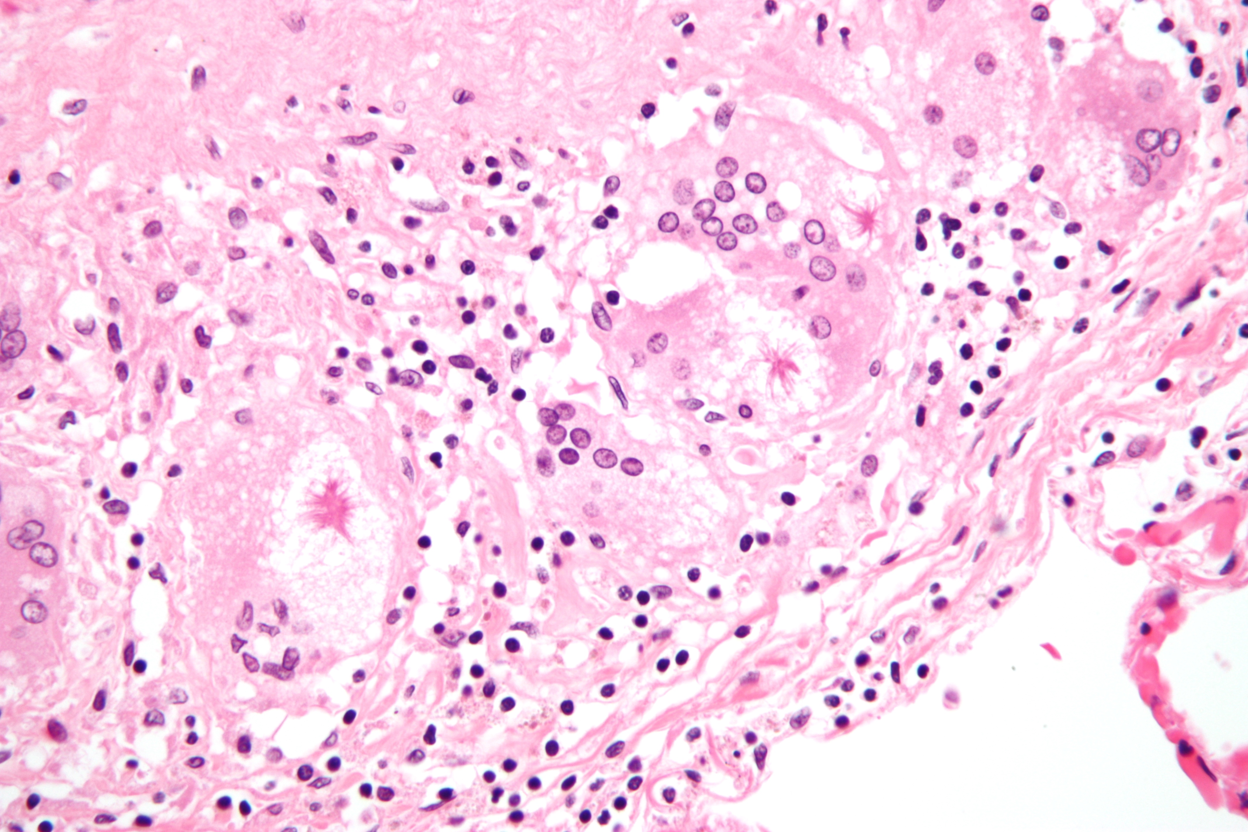
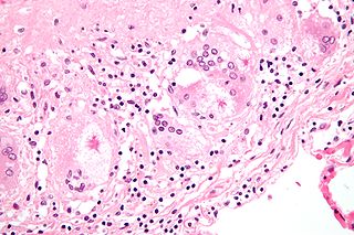
English: High magnification micrograph of asteroid bodies as seen in pulmonary sarcoidosis. H&E stain.
Asteroid bodies are cytoplasmic inclusions associated with granulomatous inflammation. On a H&E stained section, as seen here, they are light pink and have a star-fish like morphology and surrounded by white (clearing). They are suggestive of sarcoidosis, but not pathognomonic. Related images
|
| Source | Own work |
| Author | Nephron |


Licensing
[edit]
I, the copyright holder of this work, hereby publish it under the following licenses:
This file is licensed under the Creative Commons Attribution-Share Alike 3.0 Unported license.
- You are free:
- to share – to copy, distribute and transmit the work
- to remix – to adapt the work
- Under the following conditions:
- attribution – You must give appropriate credit, provide a link to the license, and indicate if changes were made. You may do so in any reasonable manner, but not in any way that suggests the licensor endorses you or your use.
- share alike – If you remix, transform, or build upon the material, you must distribute your contributions under the same or compatible license as the original.

|
Permission is granted to copy, distribute and/or modify this document under the terms of the GNU Free Documentation License, Version 1.2 or any later version published by the Free Software Foundation; with no Invariant Sections, no Front-Cover Texts, and no Back-Cover Texts. A copy of the license is included in the section entitled GNU Free Documentation License. |
You may select the license of your choice.
| Annotations | This image is annotated: View the annotations at Commons |
File history
Click on a date/time to view the file as it appeared at that time.
| Date/Time | Thumbnail | Dimensions | User | Comment | |
|---|---|---|---|---|---|
| current | 03:26, 3 September 2009 | | 4,272 × 2,848 (2.65 MB) | Nephron (talk | contribs) | {{Information |Description={{en|1=High magnification micrograph of an '''asteroid body''', as seen in pulmonary sarcoidosis. H&E stain. }} |Source=Own work by uploader |Author=[[User: |
You cannot overwrite this file.
File usage on Commons
There are no pages that use this file.
File usage on other wikis
The following other wikis use this file:
- Usage on ar.wikipedia.org
- Usage on en.wikipedia.org
- Usage on es.wikipedia.org