File:Astacus development embryo.jpg

Original file (984 × 740 pixels, file size: 349 KB, MIME type: image/jpeg)
Captions
Captions
Summary
[edit]
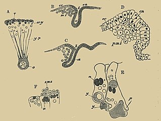
| Description | FIG. 737. FIGURES ILLUSTRATING THE DEVELOPMENT OF ASTACUS. (From Parker ; after Reichenbach.) A. Section through part of the ovum during segmentation, n. nuclei ; w.y. white yolk ; y.p. yolk pyramids ; c. central yolk mass. B and C. Longitudinal sections during the gastrula stage, a. archenteron ; b. blastopore ; ms. mesoblast ; ec. epiblast ; en. hypoblast distinguished from epiblast by shading. D. Highly magnified view of the anterior lip of blastopore to show the origin of the primary mesoblast from the wall of the archenteron. p.ms. primary mesoblast ; ec. epiblast ; en. hypoblast. E Two hypoblast cells to shew the amoeba-like absorption of yolk spheres. y. yolk ; n. nucleus ; p. pseudopodial process. F. Hypoblast cells giving rise endogenously to the secondary mesoblast (s.nts.). n. nuclei. |
| Date | |
| Source |
https://archive.org/details/theworks02balfuoft/page/510/mode/2up?view=theater&q=blastoderm THE WORKS OF FRANCIS MAITLAND BALFOUR VOL. III A TREATISE ON COMPARATIVE EMBRYOLOGY. |
| Author | M. FOSTER, F.R.S., ADAM SEDGWICK, M.A., |
Licensing
[edit]
|
This work is in the public domain in its country of origin and other countries and areas where the copyright term is the author's life plus 70 years or fewer.
| |
| This file has been identified as being free of known restrictions under copyright law, including all related and neighboring rights. | |

|
This file, which was originally posted to an external website, has not yet been reviewed by an administrator or reviewer to confirm that the above license is valid. See Category:License review needed for further instructions.
|
File history
Click on a date/time to view the file as it appeared at that time.
| Date/Time | Thumbnail | Dimensions | User | Comment | |
|---|---|---|---|---|---|
| current | 14:28, 2 March 2024 | | 984 × 740 (349 KB) | Rasbak (talk | contribs) | == {{int:filedesc}} == {{Information |description=FIG. 737. FIGURES ILLUSTRATING THE DEVELOPMENT OF ASTACUS. (From Parker ; after Reichenbach.) A. Section through part of the ovum during segmentation, n. nuclei ; w.y. white yolk ; y.p. yolk pyramids ; c. central yolk mass. B and C. Longitudinal sections during the gastrula stage, a. archenteron ; b. blastopore ; ms. mesoblast ; ec. epiblast ; en. hypoblast distinguished from epiblast by shading. I '. Highly magnified view of the anterior lip... |
You cannot overwrite this file.
File usage on Commons
There are no pages that use this file.