File:Aphis rosae segmentation.jpg

Original file (935 × 387 pixels, file size: 345 KB, MIME type: image/jpeg)
Captions
Captions
Summary
[edit]
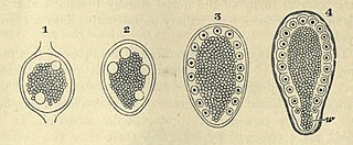
| Description | FIG. 57. SEGMENTATION OF APHIS ROSAE. (Copied from Metschnikoff.) In all the stages there is seen to be a central yolk mass surrounded by a layer of protoplasm. In this protoplasm two nuclei have appeared in 1, four nuclei in 2. In 3 the nuclei have arranged themselves regularly, and in 4 the protoplasm has become divided into a number of columnar cells corresponding to the nuclei. w. pole of the blastoderm which has no share in forming the embryo |
| Date | |
| Source |
https://archive.org/details/theworks02balfuoft/page/116/mode/2up?view=theater&q=blastoderm THE WORKS OF FRANCIS MAITLAND BALFOUR VOL. III A TREATISE ON COMPARATIVE EMBRYOLOGY. |
| Author | M. FOSTER, F.R.S., ADAM SEDGWICK, M.A., |
Licensing
[edit]
|
This work is in the public domain in its country of origin and other countries and areas where the copyright term is the author's life plus 70 years or fewer.
| |
| This file has been identified as being free of known restrictions under copyright law, including all related and neighboring rights. | |

|
This file, which was originally posted to an external website, has not yet been reviewed by an administrator or reviewer to confirm that the above license is valid. See Category:License review needed for further instructions.
|
File history
Click on a date/time to view the file as it appeared at that time.
| Date/Time | Thumbnail | Dimensions | User | Comment | |
|---|---|---|---|---|---|
| current | 22:34, 29 February 2024 | | 935 × 387 (345 KB) | Rasbak (talk | contribs) | {{Information |description=FIG. 57. SEGMENTATION OF APHIS ROSAE. (Copied from Metschnikoff.) In all the stages there is seen to be a central yolk mass surrounded by a layer of protoplasm. In this protoplasm two nuclei have appeared in 1, four nuclei in 2. In 3 the nuclei have arranged themselves regularly, and in 4 the protoplasm has become divided into a number of columnar cells corresponding to the nuclei. w. pole of the blastoderm which has no share in forming the embryo |source=https://ar... |
You cannot overwrite this file.
File usage on Commons
The following page uses this file:
- File:Aphis rosa segmentation.jpg (file redirect)
File usage on other wikis
The following other wikis use this file:
- Usage on nl.wikipedia.org