File:Anatomy of the ear, John Cunningham Saunders, 1806 Wellcome L0035337.jpg

Original file (2,922 × 4,182 pixels, file size: 4.19 MB, MIME type: image/jpeg)
Captions
Captions
Summary
[edit]
| Anatomy of the ear, John Cunningham Saunders, 1806 | |||
|---|---|---|---|
| Title |
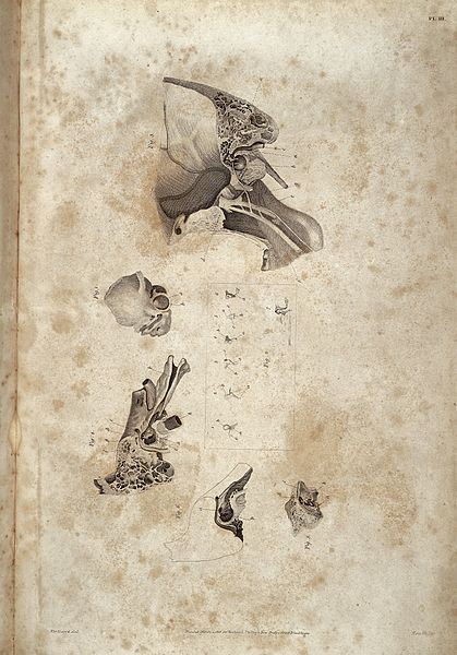
Anatomy of the ear, John Cunningham Saunders, 1806 |
||
| Description |
Anatomical illustration of the human ear showing Fig 1) Foetus Os Temporis. Fig 2) Individual bones which form chain of connection between the Membrana Tympani and Membrane of the Vestibule. Fig 3) Exterior portion of the Mastoid process and Tympanum. Fig 4) Interior portion of the Mastoid process. Fig 5) Interior Superficies of the Tympanum dissected to show Stapedeus Muscle and the Canal of Bone which lodges the Tensor Membranae Tympani. Fig 6) Skeleton of the interior superficies of the Tympanum (the Mastoid Cells being in outline) that the Fenestra Ovata and Fenestra Rotunda may be seen. Rare Books |
||
| Credit line |
|
||
| References |
|
||
| Source/Photographer |
https://wellcomeimages.org/indexplus/obf_images/22/bb/f67c1d9fa0eaad07744fd7db209c.jpg
|
||
Licensing
[edit]
- You are free:
- to share – to copy, distribute and transmit the work
- to remix – to adapt the work
- Under the following conditions:
- attribution – You must give appropriate credit, provide a link to the license, and indicate if changes were made. You may do so in any reasonable manner, but not in any way that suggests the licensor endorses you or your use.
File history
Click on a date/time to view the file as it appeared at that time.
| Date/Time | Thumbnail | Dimensions | User | Comment | |
|---|---|---|---|---|---|
| current | 07:40, 12 October 2014 | | 2,922 × 4,182 (4.19 MB) | Fæ (talk | contribs) | =={{int:filedesc}}== {{Artwork |artist = |author = |title = Anatomy of the ear, John Cunningham Saunders, 1806 |description = Anatomical illustration of the human ear showing Fig 1) Foetus Os Temporis. Fig 2)... |
You cannot overwrite this file.
File usage on Commons
The following page uses this file: