File:Anaphase eukaryotic mitosis.svg
Jump to navigation
Jump to search

Size of this PNG preview of this SVG file: 800 × 563 pixels. Other resolutions: 320 × 225 pixels | 640 × 450 pixels | 1,024 × 720 pixels | 1,280 × 900 pixels | 2,560 × 1,800 pixels | 994 × 699 pixels.
Original file (SVG file, nominally 994 × 699 pixels, file size: 146 KB)
Captions
Captions
Add a one-line explanation of what this file represents
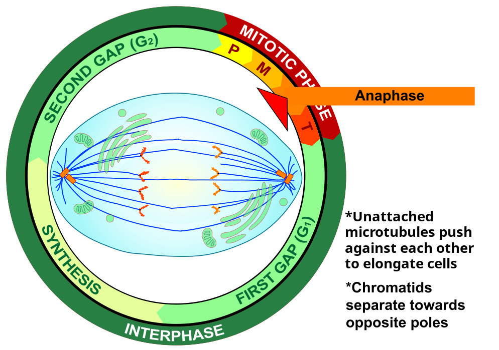
| Description | Mitosis is the process by which a eukaryotic cell separates. | ||
| Date | |||
| Source | Made myself based on the information in wikipedia, and the images:[1], [2], [3], [4] and the video:[5] | ||
| Author | LadyofHats | ||
| Permission (Reusing this file) |
|
![[1]](http://www.uhh.hawaii.edu/~ronald/392/Mitosis.JPG)
![[2]](http://www.maph49.galeon.com/mitosis/stages1.gif)
![[3]](http://www.le.ac.uk/ge/genie/vgec/images/mitosis.png)
![[4]](http://post.queensu.ca/~forsdyke/images/darwin04.gif)
File history
Click on a date/time to view the file as it appeared at that time.
| Date/Time | Thumbnail | Dimensions | User | Comment | |
|---|---|---|---|---|---|
| current | 21:49, 7 February 2018 | | 994 × 699 (146 KB) | Rojoxiii (talk | contribs) | Added text about microtubules and elongated microtubules to reflect the process. |
| 14:19, 1 December 2008 |  | 994 × 699 (129 KB) | LadyofHats (talk | contribs) | cromosomes in < > form | |
| 18:18, 8 September 2008 |  | 994 × 699 (126 KB) | LadyofHats (talk | contribs) | {{Information |Description=Mitosis is the process by which a eukaryotic cell separates. |Source=Made myself based on the information in wikipedia, and the images:[http://www.uhh.hawaii.edu/~ronald/392/Mitosis.JPG], [http://www.maph49.galeon.com/mitosis/st |
You cannot overwrite this file.
File usage on Commons
The following 3 pages use this file:
File usage on other wikis
The following other wikis use this file:
- Usage on bg.wikipedia.org
- Usage on es.wikipedia.org
- Usage on nl.wikibooks.org
- Usage on pl.wikipedia.org